Background
Athletic foot injuries can be difficult to properly diagnose and treat. Bearing the weight of the entire body, the foot is under tremendous stress. In many sports, the foot absorbs tremendous shearing and loading forces, sometimes reaching over 20 times the person's body weight. Physicians who treat these disorders must have a good understanding of the anatomy and kinesiology of the foot.
See the following images.
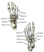 Select bones of the foot (dorsal and plantar views).
 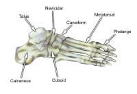 Select bones of the foot (superolateral view).
 Although foot injuries can occur from a variety of causes, the most common cause is trauma. Other etiologies include (1) rapid or improper warm-up, (2) overuse, (3) intense workouts, (4) improper footwear, and (5) playing on hard surfaces.[1, 2, 3, 4, 5]
Physicians who evaluate and treat common foot problems should have a working knowledge of the individual sports and the injuries that are commonly associated with them. An understanding of the basic treatment approaches for these injuries also is imperative.
For excellent patient education resources, visit eMedicineHealth's First Aid and Injuries Center. Also, see eMedicineHealth's patient education articles Broken Foot and Broken Toe.
NextEpidemiologyFrequencyUnited StatesEstimates indicate that 15% of sports-related injuries affect the foot alone.
PreviousNextFunctional AnatomyFoot and ankleThe foot is composed of 26 major bones, which can be divided into 3 regions: the forefoot, midfoot, and hindfoot. The forefoot is comprised of the 5 metatarsals and the 14 phalanges. The 3 cuneiforms (ie, lateral, intermediate, medial), the cuboid, and the navicular represent the midfoot. The hindfoot is composed of the talus and the calcaneus (see image below).
 The hindfoot is composed of the talus and the calcaneus. The talus is oriented to transmit forces from the foot through the ankle to the leg.The calcaneus is the largest bone in the foot. The Achilles tendon inserts on the posterior aspect of the calcaneus.The navicular lies anterior to the talus and medial to the cuboid.The cuboid articulates with the calcaneus proximally, with the fourth and fifth metatarsals distally, and with the lateral cuneiform medially (see image below).
 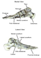 Select bones of the foot (medial and lateral views). Each of the cuneiform bones is wedge-shaped. The medial, intermediate, and lateral cuneiform bones articulate with the first 3 metatarsals distally and the navicular proximally. The cuboid articulates with the lateral cuneiform. The 5 metatarsals articulate with the proximal phalanges.The great toe is composed of 2 phalanges, with 3 for each lesser toe.Although variation exists in the number and location of the sesamoid bones, 2 constant sesamoids are present beneath the metatarsal head. The sesamoids are usually present within tendons juxtaposed to articulations.
 Select tendons of the foot (see image below)
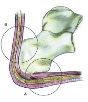
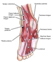 Select tendons of the foot. The flexor hallucis longus (FHL) tendon is 1 of 3 structures that lie in the tarsal tunnel. Running behind the medial malleolus, the FHL is the most posterolateral. The FHL runs anterior to insert onto the distal phalanx of the great toe. The FHL acts as a flexor of the great toe, elevates the arch, and assists with plantar flexion of the ankle. The flexor digitorum longus (FDL) tendon passes between the FHL and tibialis posterior tendon. The FDL inserts onto the distal phalanges of the 4 lateral digits and acts to flex the distal phalanges. The tibialis posterior tendon is the most anteromedial of the tarsal tunnel tendons. This tendon inserts on the navicular tuberosity; the 3 cuneiforms; the cuboid; and the second, third, and fourth metatarsals. The tibialis posterior muscle flexes, inverts, and adducts the foot. Laterally, the peroneus longus and peroneus brevis tendons share the common peroneal tunnel running behind and around the lateral malleolus. The peroneus longus plantar flexes the first metatarsal, flexes the ankle, and abducts the foot. The peroneus brevis flexes the ankle and everts the foot. Other important structuresThe plantar aponeurosis or fascia is a deep span of connective tissue extending from the anteromedial tubercle of the calcaneus to the proximal phalanges of each of the toes. Medial and lateral fibrous septa originate from the medial and lateral borders to attach to the first and fifth metatarsal bones. Nerve innervation of the foot runs along the medial and lateral metatarsals and phalanges in a neurovascular bundle. These nerves are vulnerable to compressive forces that, in time, can generate the painful Morton neuroma, which most commonly affects the interspace between the third and fourth metatarsals. Four nerves supply the forefoot: the sural nerve (most lateral), branches of the superficial peroneal nerve, the deep peroneal nerve, and the saphenous nerve. The joint between the forefoot and the midfoot, the tarsometatarsal (TMT) joint or Lisfranc joint, is formed by a mortise of the cuneiform bones surrounding the base of the second metatarsal. This joint is supported by the transverse ligaments, and the Lisfranc ligament joins the medial cuneiform and the base of the second metatarsal. Disruption of this ligament can result in a destabilization of the TMT joint complex of the foot, the result of which can be instability of the arch and the midfoot. PreviousNextSport-Specific Biomechanics
The 3 planes in which the foot and ankle function are the transverse, sagittal, and frontal. Movement is possible in all 3 planes.
Plantar flexion and dorsiflexion occur in the sagittal plane. Plantar flexion involves the foot moving from the anterior leg distally. Dorsiflexion is the opposite motion. Inversion and eversion occur in the frontal plane of motion. Eversion occurs when the bottom of the foot turns away from the midline of the body. Inversion is the opposite action. The 2 transverse plane motions are abduction and adduction. Adduction involves the foot moving toward the midline of the body, whereas abduction is the opposite action. PreviousProceed to Clinical Presentation , Athletic Foot Injuries


0 comments:
Post a Comment
Note: Only a member of this blog may post a comment.