Background
Cervical radiculopathy is a dysfunction of a nerve root of the cervical spine. The seventh (C7; 60%) and sixth (C6; 25%) cervical nerve roots are the most commonly affected.[1, 2, 3, 4, 5, 6, 7]
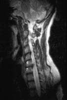 Sagittal magnetic resonance image of the cervical spine. This image reveals a C6-C7 herniated nucleus pulposus.
 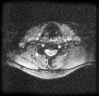 Axial magnetic resonance image of the cervical spine. This image reveals a C6-C7 herniated nucleus pulposus.
 In the younger population, cervical radiculopathy is a result of a disc herniation or an acute injury causing foraminal impingement of an exiting nerve.[8] Disc herniation accounts for 20-25% of the cases of cervical radiculopathy. In the older patient, cervical radiculopathy is often a result of foraminal narrowing from osteophyte formation, decreased disc height, degenerative changes of the uncovertebral joints anteriorly and of the facet joints posteriorly.
Factors associated with increased risk include heavy manual labor requiring the lifting of more than 25 pounds, smoking, and driving or operating vibrating equipment. Other, less frequent causes include tumors of the spine, an expanding cervical synovial cyst, synovial chondromatosis in the cervical facet joint, giant cell arteritis of the cervical radicular vessels, and spinal infections.[9, 10] The purpose of this article is to provide information on the presentation, evaluation, differential diagnosis, and treatment of cervical radiculopathy.
For excellent patient education resources, visit eMedicineHealth's First Aid and Injuries Center. Also, see eMedicineHealth's patient education articles Shoulder and Neck Pain and Neck Strain.
NextEpidemiologyFrequencyUnited StatesCervical radiculopathy occurs at a much lower frequency than radiculopathy of the lumbar spine. The annual incidence is approximately 85 cases per 100,000 population.
PreviousNextFunctional AnatomySeven cervical vertebrae and 8 cervical nerve roots exist. The C1-2 (atlantoaxial) joint forms the upper cervical segment.[1, 3, 11, 12] This joint allows for 50% of all cervical rotation. The occipitoatlantal joint is responsible for 50% of flexion and extension. Below the C2-C3 level, lateral bending of the cervical spine is coupled with rotation in the same direction. This is due to the 45° inclination of the cervical facet joints.
The vertebral bodies of C3-C7 are similar in appearance and function. They articulate via the zygapophyseal or facet joints posteriorly. On the lateral aspect of the vertebral bodies are sharply defined margins, which articulate with the facet above. These articulations are called uncovertebral joints, or the joints of Luschka. These joints can develop osteophytic spurs, which can narrow the intervertebral foramina.
Intervertebral discs are located between the vertebral bodies of C2-C7. The discs are composed of an outer annular fibrosis and an inner nucleus pulposus and serve as force dissipators, transmitting compressive loads throughout a range of motion (ROM). The intervertebral discs are thicker anteriorly and therefore contribute to normal cervical lordosis.
The foramina are largest at C2-C3 and progressively decrease in size to the C6-C7 level. The nerve root occupies 25-33% of the foraminal space. The neural foramen is bordered anteromedially by the uncovertebral joints, posterolaterally by facet joints, superiorly by the pedicle of the vertebra above, and inferiorly by the pedicle of the lower vertebra. Medially, the foramina are formed by the edge of the end plates and the intervertebral discs. The nerve roots exit above their correspondingly numbered vertebral body from C2-C7. C1 exits between the occiput and atlas, and C8 exits below the C7 vertebral body. Degenerative changes of the structures that form the foramina can cause nerve root compression. This compression can occur from osteophyte formation, disc herniation, or a combination of the 2.
PreviousNextSport-Specific BiomechanicsCervical radiculopathy in athletes can occur from several mechanisms. These injuries can occur from an extension, lateral bending, or rotation mechanism, which closes the neural foramen and results in ipsilateral nerve root injury. Conversely, a traction injury can occur with a sudden flexion or extension, coupled with lateral bending away from the affected nerve root.
Additionally, cervical disc herniations can occur with a sudden load with the neck in either flexion or extension. In elderly persons with osteophyte formation, repetitive neck extension and rotation in certain sports, such as swimming or tennis, may result in a more insidious injury.
PreviousProceed to Clinical Presentation , Cervical Radiculopathy


0 comments:
Post a Comment
Note: Only a member of this blog may post a comment.