Background
Injuries to the peroneal tendons are common but not always clinically significant.[1] They are misdiagnosed as a lateral ankle sprain most of the time, because isolated injury to the peroneal tendons is rare.[2, 3] Injury can occur in one or both peroneus longus and brevis tendons and is typically classified as acute or chronic. Function can be severely compromised by any tendon disruption; conversely, complete tendon rupture can be asymptomatic. Lesions have been seen in symptomatic patients, as well as in cadaver studies of patients who were presumably asymptomatic.[4] The reason for this variation is not known.
The image below depicts the anatomy of the lateral ankle.
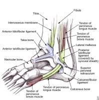 Lateral ankle anatomy demonstrates the peroneal tendons as they course beneath the superior retinaculum. The anterior talofibular, calcaneofibular, and posterior talofibular ligaments are also shown.
 Acute injuries of the peroneal tendons include tendinitis, tear/rupture, laceration, and dislocation/subluxation. Acute injuries typically have 1 of 2 mechanisms as the cause: (1) inversion ankle injury, which is often seen with associated anterior talofibular ligament and/or calcaneofibular ligament disruption, and (2) a powerful contraction of the peroneal muscles with a forcefully dorsiflexed foot.
Chronic injuries include longitudinal tears[5, 6, 7, 8, 9] and recurrent subluxation[10, 11, 12] of the peroneus brevis tendon.[13] These chronic injuries are usually associated with ankle or subtalar arthritis and ankle instability. People with "bad" or "weak" ankles may have peroneal tendon pathology. Core and lower extremity biomechanics must be evaluated in any chronic atraumatic peroneal tendinopathy, as flaws in those mechanics are usually the culprit.
For excellent patient education resources, visit eMedicineHealth's First Aid and Injuries Center. Also, see eMedicineHealth's patient education article Ankle Sprain.
NextEpidemiologyFrequencyUnited StatesThe occurrence of injuries to the peroneal tendons is not actually known. DiGiovanni et al found that 25-77% of patients with chronic lateral ankle instability had some type of injury to the peroneal tendons.[14] Over 33 months, Fallat et al noted that of 638 acute ankle "sprains" seen at the Oakwood Hospital Downriver Center Emergency Room and Occupational Medicine Clinic in Dearborn, Michigan, only 83 involved damage to the peroneal tendons, whereas more than 450 involved the anterior talofibular ligament.[1]
PreviousNextFunctional AnatomyThe peroneal tendons originate in the lateral compartment of the leg. The peroneus longus originates from the head and proximal two thirds of the fibula, whereas the peroneus brevis originates from the distal two thirds of the fibula. Both tendons have a musculotendinous portion that courses just below the lateral malleolus.
At the posterior aspect of the lateral malleolus, the peroneal tendons lie within the fibular groove, with the peroneus brevis medial and anterior to the peroneus longus. The fibular groove forms the anterior border of the fibro-osseous tunnel that the peroneal tendons course through. The inferior retinaculum and the calcaneofibular ligament form the posterior border.
The posterior talofibular and the calcaneofibular ligaments form the medial border. The superior retinaculum forms the lateral border. Just inferior to the lateral malleolus, the peroneus brevis courses anteriorly, crossing over the cuboid to insert on the fifth metatarsal styloid. (See the following image.)
Lateral ankle anatomy demonstrates the peroneal tendons as they course beneath the superior retinaculum. The anterior talofibular, calcaneofibular, and posterior talofibular ligaments are also shown. Inferior to the peroneus brevis, the peroneus longus turns beneath the cuboid in a tunnel formed by the long plantar ligament and the groove of the cuboid. It then courses to insert onto the first metatarsal and medial cuneiform. In 20% of the population, an os peroneum may be present within the peroneus longus tendon as it turns under the cuboid bone. In 0.1% of the population, a structure known as the os vesalianum—a sesamoid bone—is found at the insertion of the peroneus brevis tendon.
PreviousNextSport-Specific BiomechanicsMost sports have elements of running and lateral movement. Sports such as soccer, basketball, and football can be highly demanding on the lower extremity.
The role of the peroneus muscles is to evert the ankle and stabilize its subtalar motion. In balancing the foot, they play off the posterior tibialis muscle on the opposite side of the tibia. Maximal exertion occurs with side-to-side movement and jumping.
The importance of the peroneus muscles is most obvious after lateral ankle sprains. Trauma to the lateral ankle distorts the proprioceptive sense and stretches the connective tissues. The peroneus muscles are often stretched and injured from traction when the foot inverts.
Ankle instability ensues and continues until the lateral retinaculum heals, the peroneal muscles recover, and proprioception returns. If the retinaculum does not heal properly and cannot retain its tension to stabilize the peroneal tendons, symptoms of instability may not resolve without further intervention.
An analysis of overall biomechanics is essential in finding out the factors involved with peroneal tendon damage, especially when there is no traumatic insult. Leg-length discrepancies, femoroacetabular impingement, core instability, and low back pain are some of the correlated factors involved with lower extremity repetitive injuries, but little research has cemented the relationship. However, the core is the powerhouse of the body, and if foot planting is not well controlled by the hip and thigh, then extraneous forces run through the lower leg, ankle, and foot. This can only be controlled by increasing the activity of the supporting muscles, of which the peroneal tendons belong.
PreviousProceed to Clinical Presentation , Peroneal Tendon Syndromes


0 comments:
Post a Comment
Note: Only a member of this blog may post a comment.