Background
The spectrum of femoral shaft fractures is wide and ranges from nondisplaced femoral stress fractures to fractures associated with severe comminution and significant soft-tissue injury. Femoral shaft (see image below) fractures are generally caused by high-energy forces and are often associated with multisystem trauma. Isolated injuries can occur with repetitive stress and may occur in the presence metabolic bone diseases, metastatic disease, or primary bone tumors.[1, 2]
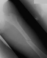 An example of an isolated, short, oblique midshaft femoral fracture, which is very amenable to intramedullary nailing. Although not seen in this x-ray film, radiographic visualization of both the proximal and distal joints should be performed for all diaphyseal fractures.
 Most femoral diaphyseal fractures are treated surgically with intramedullary nails or plate fixation. The goal of treatment is reliable anatomic stabilization, allowing mobilization as soon as possible. Surgical stabilization is also important for early extremity function, allowing both hip and knee motion and strengthening. Injuries and fractures of the femoral shaft may have significant short- and long-term effects on the hip and knee joints if alignment is not restored.
Treatment of femoral shaft fractures has undergone significant evolution over the past century. Until the recent past, the definitive method for treating femoral shaft fractures was traction or splinting. Before the evolution of modern aggressive fracture treatment and techniques, these injuries were often disabling or fatal. Traction as a treatment option has many drawbacks, including poor control of the length and alignment of the fractured bone, development of pulmonary insufficiency, deep vein thrombosis, and joint stiffness due to supine positioning.
The femur is very vascular and fractures can result in significant blood loss into the thigh. Up to 40% of isolated fractures may require transfusion, as such injuries can result in loss of up to 3 units of blood.[3] This factor is significant, especially in elderly patients who have less cardiac reserve.
Femoral fracture patterns vary according to the direction of the force applied and the quantity of force absorbed. A perpendicular force results in a transverse fracture pattern, an axial force may injure the hip or knee, and rotational forces may cause spiral or oblique fracture patterns. The amount of comminution present increases with the amount of energy absorbed by the femur at the time of fracture.[1, 2, 4, 5]
For excellent patient education resources, visit eMedicineHealth's First Aid and Injuries Center. Also, see eMedicineHealth's patient education article Broken Leg.
Related Medscape Reference topics:
Femoral Neck Stress and Insufficiency Fractures [in the Orthopedic Surgery section]
Femoral Neck Stress Fracture
Femur Fracture [in the Emergency Medicine section]
Related Medscape resources:
Resource Center Exercise and Sports Medicine
Specialty Site Emergency Medicine
Specialty Site Orthopaedics
CMEÂ A 49-Year-Old Man With a Femur Fracture and Hyperdense Bones
CMEÂ Vitamin D and Musculoskeletal Health
NextEpidemiologyFrequencyUnited StatesThe incidence of femoral fractures is reported as 1-1.33 fractures per 10,000 population per year (1 case per 10,000 population). In individuals younger than 25 years and those older than 65 years, the rate of femoral fractures is 3 fractures per 10,000 population annually. These injuries are most common in males younger than 30 years. Causes may include automobile, motorcycle, or recreational vehicle accidents or gunshot wounds. The average number of days lost from work or school from femoral fractures is 30.The average number of days of restricted activity due to femoral fractures is 107.The incidence of femoral injuries and fractures increases in elderly patients.PreviousNextFunctional AnatomyThe femur is the strongest, longest, and heaviest bone in the body and is essential for normal ambulation. It consists of 3 parts (ie, femoral shaft or diaphysis, proximal metaphysis, distal metaphysis). The femoral shaft is tubular with a slight anterior bow, extending from the lesser trochanter to the flare of the femoral condyles. During weight bearing, the anterior bow produces compression forces on the medial side and tensile forces on the lateral side. The femur is a structure for standing and walking, and it is subject to many forces during walking, including axial loading, bending, and torsional forces. During contraction, the large muscles surrounding the femur account for most of the applied forces.[1, 2, 4, 5]
Several large muscles attach to the femur. Proximally, the gluteus medius and minimus attach to the greater trochanter, resulting in abduction of the femur with fracture. The iliopsoas attaches to the lesser trochanter, resulting in internal rotation and external rotation with fractures. The linea aspera (rough line on the posterior shaft of the femur) reinforces the strength and is an attachment for the gluteus maximus, adductor magnus, adductor brevis, vastus lateralis, vastus medialis, vastus intermedius, and short head of the biceps. Distally, the large adductor muscle mass attaches medially, resulting in an apex lateral deformity with fractures. The medial and lateral heads of the gastrocnemius attach over the posterior femoral condyles, resulting in flexion deformity in distal-third fractures.
The blood supply enters the femur through metaphyseal arteries and branches of the profunda femoris artery, penetrating the diaphysis and forming medullary arteries extending proximally and distally. With intramedullary nailing, the blood supply is disrupted and progressively reestablishes itself over 6-8 weeks. Healing of the fracture is enhanced by the surrounding soft tissue and local recruitment of blood supply around the callus. The femoral artery courses down the medial aspect of the thigh to the adductor hiatus, at which time it becomes the popliteal artery. Injuries to the artery occur at the level of the adductor hiatus, where soft-tissue attachments may cause tethering. Uncommonly, the sciatic nerve is injured in femoral shaft fractures; however, it may become injured in proximal or distal femoral injuries.
Related Medscape Reference topics:
Nerve Entrapment Syndromes [in the Neurosurgery section]
Nerve Entrapment Syndromes of the Lower Extremity [in the Orthopedic Surgery section]
PreviousNextSport-Specific BiomechanicsTrauma-induced fractures of the femur occur with contact and during high-speed sports. A significant amount of energy is transferred to the limb in a femur fracture, such as might be generated in skiing, football, hockey, rodeo, and motor sports.
Stress fractureA femoral stress fracture is the result of cyclic overloading of the bone or a dramatic increase in the muscular forces across their insertion, causing microfracture. These repetitive stresses overcome the ability of the bone to heal the microtrauma. The area most susceptible to stress fracture is the medial junction of the proximal and middle third of the femur, which occurs as a result of the compression forces on the medial femur.
Stress fractures can also occur on the lateral aspect of the femoral neck in areas of distraction and are less likely to heal nonoperatively than compression-side stress fractures. Stress fractures occur most often in repetitive overload sports such as in runners and in baseball and basketball players. For more information, refer to the Medscape Reference article Femoral Neck Stress Fracture.
PreviousProceed to Clinical Presentation , Femur Injuries and Fractures


0 comments:
Post a Comment
Note: Only a member of this blog may post a comment.