Background
Sprains and dislocations of the metacarpophalangeal (MCP) joint of the finger are relatively rare due to the protected position of this joint in the hand.[1, 2, 3, 4] Injuries to the MCP joint of the thumb are more common, although these usually consist of collateral ligament injuries rather than dorsal or palmar dislocations.[5, 6]
MCP joint dislocation is seen in the image below.
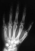 Metacarpophalangeal joint dislocation of the small finger. Posteroanterior radiograph demonstrates loss of joint space.
 For patient education resources, see the Bone, Joint, and Muscle Center and Breaks, Fractures, and Dislocations Center, as well as Finger Dislocation, Broken Finger, and Sprains and Strains.
NextFunctional AnatomyThe bony anatomy of the finger MCP joint provides greater laxity in extension, with the shallow articular surface of the proximal phalanx resting on the spherical metacarpal head. The metacarpal head is wider in palmar orientation, which leads to increasing bony stability as the joint approaches maximal flexion. Soft-tissue constraints, including the volar plate, accessory and true collateral ligaments, dorsal capsule, extensor tendon and sagittal band, and intrinsic tendons provide additional stability to the MCP joint. This results in an arc of motion from 30º of hyperextension to 120º of flexion, 30-40º of mediolateral laxity, and a small degree of rotational laxity.
The volar plate is a fibrocartilaginous structure firmly attached to the base of the proximal phalanx. Its origin, just proximal to the metacarpal head, is thin and diaphanous; this allows hyperextension of the MCP joint, but it is also the part of the joint most susceptible to injury during dislocations. The deep transverse metacarpal ligaments further stabilize the volar plates of the neighboring MCP joint.
The collateral ligaments originate from mediolateral depressions in the metacarpal head and travel in a distal-palmar direction to insert onto the base of the proximal phalanx. The elliptical shape of the metacarpal head causes these ligaments to loosen in extension and tighten in flexion. The accessory collateral ligament spans from the true collateral ligament to the volar plate, providing additional joint stability in extension. The central extensor tendon and sagittal band augment the thin dorsal capsule. The tendons of the palmar and dorsal interossei add a small degree of dynamic stability.
The MCP joint of the thumb is a condyloid (hinged) joint, with a quadrilateral rather than spherical metacarpal head. The capsule and ligaments of this joint are similar to those of the finger MCP joint. Additionally, the volar plate of the thumb MPJ usually contains 2 sesamoids that articulate with the metacarpal head. The insertion of the thenar muscles into the sesamoids contributes to joint stability. These bony and ligamentous constraints allow less motion than in the MCP joint of the fingers, especially in lateral motion and rotation; abduction and adduction average 10º and a slight amount of pronation occurs during flexion.
PreviousNextSport-Specific BiomechanicsDorsal MCP joint dislocations have been described as simple or complex. Simple dislocations are those in which no soft tissue is interposed in the joint. These are usually reduced easily with an appropriate closed technique. In a classic article published in 1957, Kaplan elegantly described the anatomic features of the complex MCP joint dislocation.[7] A metacarpal head displaced in palmar orientation sits between the lumbrical muscle radially and the flexor tendons ulnarly. The volar plate, still firmly attached to the base of the proximal phalanx, is displaced into the MCP joint. Longitudinal traction only further tightens these already taut soft tissues, trapping the metacarpal head. Complex dislocations usually require open reduction.
PreviousProceed to Clinical Presentation , Metacarpophalangeal Joint Dislocation


0 comments:
Post a Comment
Note: Only a member of this blog may post a comment.