Background
Voluntary, or skeletal, muscle is the largest single organ of the human body and accounts for nearly 50% of the body's weight. The number of muscles in the body depends on the degree of subdivision that is considered and on the number of variable muscles that are included. Not counting heads, bellies, and other divisions of muscles, the Nomina Anatomica reported by the International Anatomical Nomenclature Committee under the Berne Convention lists 200 paired muscles, or a total of 400 muscles. Any one of these muscles can develop myofascial trigger points (MTrPs).[1] MTrPs are hyperirritable tender spots in palpable tense bands of skeletal muscle that refer pain and motor dysfunction, often to another location.[2, 3]
The myofascial pain syndromes (MPS) owe their ever-widening acceptance to the pioneering work of Travell and her later collaboration with Simons.[2, 3] In 1983, they combined their clinical experience in a detailed description of the multiple pain syndromes attributed to this disorder. In doing so, they further defined the major clinical components that are characteristic of myofascial pain, the most important being the TrP, the taut band, and the local twitch response. See the image below.
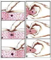 Myofascial pain in athletes. Cross-sectional drawing shows flat palpation of a taut band and its trigger point.Left: Skin pushed to one side to begin palpation (A). The fingertip slides across muscle fibers to feel the cord-line texture of the taut band rolling beneath it (B). The skin is pushed to other side at completion of movement. This same movement performed vigorously is snapping palpation (C).Right: Muscle fibers surrounded by the thumb and fingers in a pincer grip (A). The hardness of the taut band is felt clearly as it is rolled between the digits (B). The palpable edge of the taut band is sharply defined as it escapes from between the fingertips, often with a local twitch response (C). NextEpidemiologyFrequencyUnited States
MTrPs are extremely common and become a painful part of nearly everyone's life at one time or another. Latent TrPs, which often cause motor dysfunction (eg, stiffness, restricted range of motion) without pain, are far more common than active TrPs that cause pain.
Active TrPs are commonly found in postural muscles of the neck, shoulder, and pelvic girdles and in the masticatory muscles. In addition, the upper trapezius, scalene, sternocleidomastoid, levator scapulae, and quadratus lumborum muscles are commonly involved.
Reports of the prevalence of MTrPs in specific patient populations are available. The data indicate a high prevalence of this condition among individuals with a regional pain complaint, as shown in Table 1.
Table 1. Prevalence of Myofascial Pain (Open Table in a new window)
RegionPracticeNumber StudiedPrevalence of Myofascial Pain, %GeneralMedical17230GeneralPain medical center9693GeneralComprehensive pain center28385CraniofacialHead and neck pain clinic16455LumboglutealOrthopedic clinic9721The wide range in the prevalences of myofascial pain caused by TrPs is likely due to differences in the patient populations examined and in the degree of chronicity, at least in part. Probably even more important are differences in the criteria used to diagnose MTrPs and, most important, differences in the training and skill of the examiners.
PreviousNextFunctional AnatomySome isolated large round muscle fibers and some groups of these darkly staining, enlarged, round muscle fibers appear in cross-sections. In longitudinal sections, the corresponding feature is a number of contraction knots. An individual knot appears as a segment of muscle fiber with extremely contracted sarcomeres. This contractured segment has a corresponding increase in diameter of the muscle fiber.
The structural features of contraction knots presents a likely explanation for the palpable nodules and the taut bands associated with TrPs. Three single contraction knots can be seen scattered among normal muscle fibers. Beyond the thickened segment of the contractured muscle fiber at the contraction knot, the muscle fiber becomes markedly thinned and consists of stretched sarcomeres to compensate for the contractured ones in the knot segment. In addition, a pair of contraction knots separated by empty sarcolemma may represent one of the first irreversible complications that result from the continued presence of the contraction knot.
PreviousNextSport Specific BiomechanicsThe activation of a TrP is usually associated with some degree of mechanical abuse of the muscle in the form of muscle overload, which may be acute, sustained, and/or repetitive. In addition, leaving the muscle in a shortened position can convert a latent TrP to an active TrP; this process is greatly aggravated if the muscle is contracted while in the shortened position.
In paraspinal muscles (and likely other muscles, too), a degree of nerve compression that causes identifiable neuropathic electromyographic (EMG) changes is associated with an increase in the numbers of active TrPs. These TrPs may be activated by disturbed microtubular communication between the neuron and the endplate because the motor endplate is involved in the pathophysiologic process of the peripheral core TrP.
The histopathologic complications that could contribute to the chronicity of the condition and make treatment more difficult include the following:
Distortion of the striations (sarcomere arrangement) in adjacent muscle fibers for some distance beyond the contraction knot (see the image below). This produces unnatural shear forces between fibers that could seriously and chronically stress the sarcolemma of the adjacent muscle fibers. If the membrane were stressed to the point at which it became pervious to the relatively high concentration of calcium in the extracellular space, it could induce massive contracture that could compound the shear forces.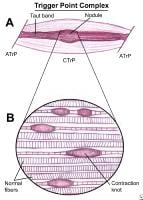 Myofascial pain in athletes. Schematic of a trigger point complex of a muscle in longitudinal section.A: The central trigger point (CTrP) in the endplate zone contains numerous electrically active loci and numerous contraction knots. A taut band of muscle fibers extends from the trigger point to the attachment at each end of the involved fibers. The sustained tension that the taut band exerts on the attachment tissues can induce a localized enthesopathy that is identified as an attachment trigger point (ATrP).B: Enlarged view of part of the CTrP shows the distribution of 5 contraction knots. The vertical lines in each muscle fiber identify the relative spacing of its striations. The space between 2 striations corresponds to the length of one sarcomere. The sarcomeres within one of these enlarged segments (ie, contraction knot) of a muscle fiber are markedly shorter and wider than the sarcomeres in the neighboring normal muscle fibers, which are free of contraction knots. The occasional finding of a segment of an empty sarcolemmal tube between 2 contractions knots may represent an additional irreversible complication of a contraction knot.
 Latent TrPs can produce other effects characteristic of a TrP, including increased muscle tension and muscle shortening; but these do not produce spontaneous pain. Both active and latent TrPs can cause significant motor dysfunction. The same factors that are responsible for the development of an active TrP can, to a lesser extent, cause a latent TrP. An active key TrP in one muscle can induce an active satellite TrP in another. Inactivation of the key TrP often inactivates its satellite TrP without treatment of the satellite TrP itself.
The intensity and extent of the pattern of referred pain depends on the degree of irritability in the TrP, not on the size of the muscle. MTrPs in small, obscure, or variable muscles can be as troublesome to the patient as TrPs in large familiar muscles.
TrPs are activated directly by acute overload, overwork fatigue, direct impact trauma, and radiculopathy. TrPs can be activated indirectly by other existing TrPs, visceral disease, arthritic joints, joint dysfunctions, and emotional distress. Satellite TrPs are prone to develop in muscles that lie within the pain reference zone of key MTrPs or within the zone of pain referred from a diseased viscus, such as the pain due to myocardial infarction, gastric ulcer, cholelithiasis, or renal colic. A perpetuating factor increases the likelihood of overload stress that can convert a latent TrP to an active TrP.
With adequate rest and in the absence of perpetuating factors, an active TrP may spontaneously revert to a latent state. Pain symptoms disappear; however, occasional reactivation of the TrP by exceeding that muscle’s stress tolerance can account for a history of recurrent episodes of the same pain over a period of years.
PreviousProceed to Clinical Presentation , Myofascial Pain in Athletes


0 comments:
Post a Comment
Note: Only a member of this blog may post a comment.