Background
Lumbosacral spondylolysis (lumbar spondylolysis) is a unilateral or bilateral defect of the pars interarticularis that affects one or more of the lumbar vertebrae. See the images below.
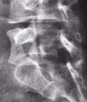 Radiograph of L4 defect in the pars interarticularis.
 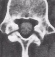 Computed tomography scan demonstrating defects in the left and right pars interarticularis.
 The term spondylolysis is derived from the Greek words spondylos, meaning vertebra, and lysis, meaning break or defect. Numerous hypotheses have been proposed on the etiology of lumbosacral spondylolysis (lumbar spondylolysis), as follows:
Separate ossification centersFracture during postnatal lifeStress fracture[1, 2] Increased lumbar lordosisImpingement of the articular process on the pars articularisWeakness of supporting structures[3] Growth[2, 4] Pathologic changes in the pars articularisDysplasia of the pars interarticularisHowever, mechanical factors are widely believed to be the cause or at least the trigger of the development of lumbosacral spondylolysis (lumbar spondylolysis), especially when congenital abnormalities are present.[5] Moreover, lumbosacral spondylolysis (lumbar spondylolysis) is argued to be related to the human erect posture and lumbar curve.[1]
Ambulation may have a role in the genesis of lumbosacral spondylolysis (lumbar spondylolysis) because no known cases exist in nonambulatory patients.[6] As an acquired condition, no reports exist of its occurrence in stillborn fetuses or in the newborn.[7] Heredity is also implicated.[8]
When the defect in the pars interarticularis is not associated with a forward displacement, the term spondylolysis applies.[9] The term spondylolisthesis is derived from spondylos and listhesis, meaning movement or slipping, and refers to the slipping forward of one vertebra on the next caudal vertebra (see the image below).
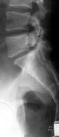 Lateral radiograph of the lumbar spine shows spondylolysis at L5 with spondylolisthesis at L5 through S1. On this single view, it is not possible to determine if these pars defects are unilateral or bilateral. Oblique views may help resolve this issue.
 Lumbosacral spondylolysis (lumbar spondylolysis) is most common at L5, accounting for 85% of all cases,[10] and may be observed as high as L2.[11] Therefore, a slip is most common at the level of L5 slipping forward on S1. Lumbosacral spondylolysis (lumbar spondylolysis) is the cause of the most common type of spondylolisthesis.[4] Moreover, Ariyoshi et al reported a case of lumbosacral spondylolysis (lumbar spondylolysis) that occurred at 3 sites in L5 that involved the bilateral pars interarticularis and the center of the right lamina.[12]
For excellent patient education resources, see eMedicineHealth's patient education article Low Back Pain.
NextEpidemiologyFrequencyUnited StatesLumbosacral spondylolysis (lumbar spondylolysis) is more commonly observed in males,[10] but this difference may not be significant.[13, 14]
In the United States, a reported difference exists between the sexes and races, with an incidence of lumbosacral spondylolysis (lumbar spondylolysis) of 6.4% in white men, 2.8% in black men, 2.3% in white women, and 1.1% in black women. A pars defect is twice as common in boys than in girls, although high-grade slippage is 4 times more common in girls than in boys. Alaskan Eskimos (26%) have the highest incidence, with the highest rate in Eskimos from north of the Yukon River.[4]
PreviousNextFunctional AnatomyRepetitive axial loading, especially in an extended lumbar spine is thought to be the most important contributing mechanism causing lumbosacral spondylolysis (lumbar spondylolysis), leading to fatigue fracture of the pars interarticularis. Shear stresses on the isthmic pars are greater when the lumbar spine is extended. When repetitive extension stresses occur, the pars interarticularis becomes impinged from the inferior facet of the cephalad vertebrae, which results in microfractures and attempts at repair.[15] See the images below.
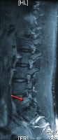 Long TR (T2-weighted) fat suppressed sagittal magnetic resonance image shows increased signal in the pars interarticularis on the left at L5 (same patient in Images 3-4). This is an acute stress reaction.
 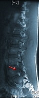 Sagittal short TR (T1-weighted) magnetic resonance image shows decreased signal in the pars interarticularis on the left at L5 (same patient in Images 3-4). PreviousNextSport-Specific Biomechanics
Lumbosacral spondylolysis (lumbar spondylolysis) occurs in 3-7% of the general population[14] The athletic population is believed to be more prone to the development of this condition,[15] because the incidence of lumbosacral spondylolysis (lumbar spondylolysis) in competitive athletes is higher than the percentage reported for the nonsports population.[16]
The overall percentage of lumbosacral spondylolysis (lumbar spondylolysis) among athletes in a study by Soler et al was about 8%, a figure not significantly higher than that among the general population.[14] However, certain sporting events were found to contribute higher percentages when each sport was considered separately, with the highest percentages of lumbosacral spondylolysis (lumbar spondylolysis) occurring in throwing sports (26.67%), artistic gymnastics (16.96%), and rowing (16.88%).[14] In an earlier series, a high percentage of lumbosacral spondylolysis (lumbar spondylolysis) was been observed in diving (43.13%), wrestling (29.82%), and weight lifting (22.68%).[16]
Other sports with high incidence rates of lumbosacral spondylolysis (lumbar spondylolysis) are ballet, dancing, football, volleyball, and fast bowlers in cricket. In ballet, the higher incidence rate is due in part to an inability to reach or maintain proper turn-out and thus overcompensation with lordosis.
In general, the presence of the repetitive actions of flexion, extension, rotation, and torsion, either alone or in combination, that are often associated with resistance are the biomechanical movements that show the highest prevalence of lumbosacral spondylolysis (lumbar spondylolysis).[14]
PreviousProceed to Clinical Presentation , Lumbosacral Spondylolysis


0 comments:
Post a Comment
Note: Only a member of this blog may post a comment.