Background
Hip and pelvis injuries represent 2-5% of all sports injuries. Among these injuries, groin pain is the most common finding. The most common sports-related injuries in the hip, pelvis, and thigh area are musculotendinous, (eg, quadriceps strain, adductor tendinitis) and, less commonly, iliopsoas tendinitis. Iliopsoas tendinitis and iliopsoas bursitis are closely interrelated because inflammation of one inevitably causes inflammation of the other, due to their close proximity. Therefore, these 2 conditions are essentially identical in terms of presentation and management.
In basic terms, iliopsoas tendonitis is an inflammation of the tendon or area surrounding the tendon. Major causes of iliopsoas tendinitis are acute trauma and overuse resulting from repetitive hip flexion. See the image below.
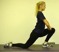 Iliopsoas stretch. NextEpidemiologyFrequencyUnited States
No data on prevalence of iliopsoas tendinitis exists. Despite this, it is a relatively uncommon and poorly recognized cause of anterior hip or groin pain. Iliopsoas tendinitis is noted to affect young adults more commonly, with a slight female predominance.
PreviousNextFunctional AnatomyThe pelvis links the trunk and lower extremities. The hip, a ball and socket joint, allows for 3 degrees of freedom. Range of motion (ROM) of the hip includes approximately 120° of flexion, 20° of extension, 40° of abduction, 25° of adduction, and 45° each of internal rotation and external rotation. The resting position of the hip is considered to be 30° of flexion and 30° of abduction.
The psoas and iliacus muscles originate from the lumbar spine and pelvis, respectively, and are innervated by the upper lumbar nerve roots (ie, L1, L2, L3). These muscles converge to form the iliopsoas muscle, which inserts onto the lesser trochanter of the proximal femur as the iliopsoas tendon. The psoas major tendon exhibits a characteristic rotation through its course, transforming its ventral surface into a medial surface. The iliac portion of this tendon has a more lateral position, and the most lateral muscle fibers of the iliacus muscle insert onto the lesser trochanter without joining the main tendon.
The iliopsoas muscle passes anterior to the pelvic brim and hip capsule in a groove between the anterior inferior iliac spine laterally and iliopectineal eminence medially. The musculotendinous junction is consistently found at the level of this groove. The iliopsoas muscle functions as a hip flexor and external rotator of the femur.
An ilio-infratrochanteric muscular bundle has been described, which likely relates to the iliopsoas tendon. This muscular bundle arises from the interspinous incisure and anterior inferior iliac spine (above the origin of the rectus femoris muscle), courses along the anterolateral edge of the iliacus muscle, and inserts without a tendon onto the anterior surface of the lesser trochanter. The iliopsoas bursa lies between the musculotendinous junction and the pelvic brim. This bursa is the largest in the body and may extend proximally into the iliac fossa or distally to the lesser trochanter. Communication between this bursa and the hip joint occurs in approximately 15% of all adults.
A variety of terms have been used to describe and classify tendon injuries. Tendonitis is typically associated with an acute injury through which failure of the tendon fibers and disruption of the vascularized peritendinous connective tissue produces an acute inflammatory response within the tendon. Tendinitis may be acute, subacute, or chronic, depending on the duration of symptoms.
Peritendinitis is a condition in which an acute injury produces an inflammatory response in only the soft tissue surrounding a tendon, without disruption of the tendon fibers. On the other hand, tendinosis is often associated with chronic microtrauma to the tendon, such as repetitive overload. In the case of tendinosis, fiber failure tends to be characterized by intrasubstance failure, compared with peritendinous disruption, which occurs in tendinitis. Microscopic findings in tendinosis include fibrillar degeneration, angiofibroblastic proliferation, myxoid degeneration, fibrosis, and, occasionally, chronic inflammation.
PreviousNextSport-Specific BiomechanicsAcute injury and overuse injury are the 2 main causes of iliopsoas tendinitis. The acute injury typically involves an eccentric contraction of the iliopsoas muscle, but also may be due to direct trauma. Overuse injury may occur in activities involving repeated hip flexion or external rotation of the thigh. Activities that may predispose to iliopsoas tendinitis include dancing, ballet, resistance training, rowing, running (particularly uphill), track and field, soccer, and gymnastics.
During the adolescent growth spurt, the hip flexors tend to become relatively inflexible. This inflexibility can lead to problems in younger athletes because stress placed on the iliopsoas musculotendinous unit increases and general biomechanics are altered. Tightness of the iliopsoas, tensor fascia lata, or rectus femoris can lead to inhibition of the gluteus maximus, allowing for an anterior pelvic tilt. This in turn leads to adverse affects on the kinetic chain. Excessive anterior tilt can lead to increased lumbar lordosis with resultant stress on the lower lumbar discs, facet joints, and sacroiliac joints and may result in increased knee flexion at heel strike and during midstance phases of the gait cycle. The subsequent increase in eccentric load across the knee extensor mechanism may result in patellar tendon injuries. With increased knee flexion, compressive forces at the patellofemoral contact surface increase and may predispose to patellofemoral problems.
PreviousProceed to Clinical Presentation , Iliopsoas Tendinitis


0 comments:
Post a Comment
Note: Only a member of this blog may post a comment.