Background
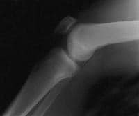
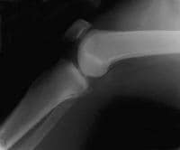
The posterior cruciate ligament (PCL) is described as the primary stabilizer of the knee by many authors. PCL injuries are less common than anterior cruciate ligament (ACL) injuries, and they often go unrecognized. The PCL is broader and stronger than the ACL and has a tensile strength of 2000 N. Injury most often occurs when a force is applied to the anterior aspect of the proximal tibia when the knee is flexed. Hyperextension and rotational or varus/valgus stress mechanisms also may be responsible for PCL tears. Injuries may be isolated or combined with other ligamentous injuries. A PCL tear can result in varying degrees of disability, from no impairment to severe impairment. PCL injury has been overly simplified, and the functional disability of PCL injury may be underestimated.[1] The radiographs belowdemonstrate the results of suchinjuries, comparing a normal knee with one that has a damaged PCL.
 A normal lateral radiograph of a knee. In a normal knee, a line drawn along the posterior femoral condyle will not intersect the posterior tibial condyle.
  A lateral radiograph of a knee with a posterior cruciate ligament injury. Note that the same line as in the above image will bisect the posterior tibial condyle due to a posterior sag and an incompetent posterior cruciate ligament.
 The primary function of the PCL is to prevent posterior translation of the tibia on the femur. The PCL also plays a role as a central axis controlling and imparting rotational stability to the knee. This injury has received little attention in the past, compared with the ACL; however, this emphasis on the ACL has stimulated increased interest in the treatment of PCL injuries. Controversy regarding treatment of isolated PCL injuries exists in the literature, with recommendations supporting both operative and nonoperative therapy. Current management of PCL injuries unfortunately can yield relatively poor clinical outcomes, whether surgically or conservatively treated.[2]
NextEpidemiologyFrequencyUnited StatesTrue incidence in the United States is unknown. In National Football League predraft physical examinations, a 2% incidence of isolated, asymptomatic, and unknown PCL injuries was found; operated, isolated, and combined PCL injuries were reported at an incidence of 3.5-20%. On the KT-1000 stress test examination, a 7% incidence of PCL injuries was found, of which 40% were isolated and unidirectional and 60% were multidirectional.
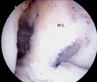
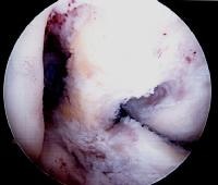
PreviousNextFunctional AnatomyAs demonstrated in the images below, the PCL originates from the intercondylar notch of the femur on the roof of the medial femoral condyle. The insertion is central on the posterior aspect of the tibial plateau, on a depression between the tibial plateaus, extending 1 cm below the articular surface.[3] The ligament is composed of a larger anterolateral bundle and a smaller posteromedial bundle. The anterior component is tightest in the midarc of flexion and the posterior fibers are tight in extension and deep flexion.
 A view of the broad origin of the posterior cruciate ligament (PCL) on the medial femoral condyle of a left knee. The anterior cruciate ligament has been removed for surgical reconstruction.
  An additional view of the posterior cruciate ligament broad origin and insertion in a knee pending anterior cruciate ligament reconstruction.
 In addition, variable anterior and posterior meniscofemoral ligaments of Humphrey and Wrisberg attach distally and proximally to the PCL, respectively. The meniscofemoral ligaments attach distally to the posterior horn of the lateral meniscus, in a slanting orientation, providing resistance to the tibial posterior drawer.[4] The PCL is an extrasynovial structure that lies behind the intra-articular portion of the knee. The primary function of the PCL is to resist posterior displacement of the tibia in relation to the femur; its secondary function is to prevent hyperextension and limit internal and varus/valgus rotation.
PreviousNextSport Specific BiomechanicsDisruption may occur with forced hyperextension while the foot is planted in dorsiflexion. A force applied to the anteromedial aspect of the knee, as during a football tackle, results in a posteriorly directed force and a varus hyperextension force, leading to PCL and posterolateral capsular ruptures.
PreviousProceed to Clinical Presentation , Posterior Cruciate Ligament Injury


0 comments:
Post a Comment
Note: Only a member of this blog may post a comment.