Background
Cervical intervertebral disc disease accounts for 36% of all spinal intervertebral disc disease, second only to lumbar disc disease, which accounts for 62% of all spinal intervertebral disc disease. Cervical problems tend to be less debilitating than lumbar problems, and they do not cause individuals to miss work as often as lumbar spine problems do.[1, 2]
One of 5 visits to an orthopedic practice is for cervical discogenic pain (CDP), with C5-6 and C6-7 accounting for approximately 75% of visits. C7 is the most common nerve root involved.[3] Cervical discogenic pain syndrome (CDPS) presents with proximal symptoms first, and, later, it can progress to brachialgia.
For excellent patient education resources, visit eMedicineHealth's First Aid and Injuries Center. Also, see eMedicineHealth's patient education articles Shoulder and Neck Pain and Neck Strain.
NextEpidemiologyFrequencyUnited StatesCervical intervertebral disc disease accounts for 36% of all spinal intervertebral disc disease. This condition is somewhat more common in women. Although acute attacks may start at a very young age with episodes of acute torticollis or "wry neck," the incidence peaks when persons are aged 45-50 years (see image below).
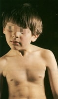 Appearance of torticollis as a result of sternomastoid fibrosis in a young child.
 Of all sports-related injuries, 2-3% are spinal injuries and the majority of these happened during unsupervised activities such as football, soccer, wrestling, diving, surfing, skiing and sand lot games.[4] The majority of the available literature, however, is found for football and this group is the most likely to sustain cervical trauma.
Statistical estimates of the incidence of cervical injury for football players varies ranging from 1 quadriplegic injury per 7,000 to 1 injury per 58,000.[5] Another review reported that since 1977, there has been an annual incidence of fewer than 10 cases of permanent injury to the cervical spinal cord among football players.[6] In 1976, the National Collegiate Athletic Association football rules committee disallowed the technique of spear tackling or the technique of using the helmet to butt or ram an opponent. This resulted in a remarkable decrease in the incidence of catastrophic neck injuries over the next 9 years.[7]
PreviousNextFunctional AnatomyThe cervical spine permits a wide range of motion (ROM) of the head in relation to the trunk. A degree of stability and flexibility is required to control the motion and dissipate the forces applied to the spine. Great differences in anatomy and function exist between the occiput-C1, the C1-2 (upper complex), and C3-C7 (lower complex) levels. Eight motion segments occur between the occiput and T1. No disc exists between C1 and C2; therefore, the first intervertebral disc is between C2 and C3.
The intervertebral disc consists of an outer annulus fibrosus and an inner nucleus pulposus. The intervertebral disc is thicker anteriorly, contributing to the normal cervical lordosis. The C6-7 disc is the thickest disc of the cervical spine. The nucleus pulposus and the inner one half of the annulus fibrosus are avascular and receive nutrition through diffusion, compression, dehydration, and imbibition of fluids.[8]
The annulus fibrosus, particularly the outer one third, has been found to be innervated by the sinuvertebral nerve and the vertebral nerve. The sinuvertebral nerve arises from the ventral ramus (somatic root), whereas the vertebral nerve (autonomic root) is derived primarily from the sympathetic nervous system. However, the vertebral nerve has connections with the cervical ventral rami, which suggests the possibility of the vertebral nerve also conveying somatic afferents from the disc.[9, 10, 11]
The nociceptors and mechanoreceptors in the annulus fibrosus mediate pain transmission from structural disruption of the intervertebral disc itself or from the chemically mediated inflammatory effect of phospholipase A2.[10, 12] Pacinian corpuscles and Golgi tendon organs present in the posterolateral region of the outer one third of the annulus transmit proprioceptive information from the intervertebral disc.[8, 12, 13, 14, 15]
The adult cervical disc has a crescentic shape anteriorly, with the apex of the crescent at the uncovertebral joints on each side. The posterior annulus has multiple vertical fissures allowing for a very degenerative appearance during discography and on gross examination. In addition, the nucleus of the cervical disc tends to be poorly centralized when compared with the lumbar disc. In the lumbar disc, the nucleus tends to be well localized in the center of the disc, and the posterior annulus tends to remain relatively intact when compared with the cervical disc. Annular fissures in the lumbar disc tend to be circumferential and/or radial in nature.
PreviousNextSport-Specific BiomechanicsBiomechanics is the study of the changes in the anatomic structures occurring during body movements. The movements of the cervical spine include flexion and extension in the sagittal plane, lateral flexion in the coronal plane, and rotation in the horizontal plane. Lateral flexion and rotation occur as coupled movements. Other movements of the cervical spine include protrusion (ie, the head is moved as far forward as possible with the neck outstretched and maintaining forward-facing position) and retraction (ie, the head is moved as far backward as possible and maintaining a forward-facing position).
Fifty percent of rotation of the cervical spine occurs in the upper cervical complex with the atlas rotating ipsilaterally around the odontoid. Protrusion causes upper cervical spine extension and lower cervical spine flexion, whereas retraction causes upper cervical spine flexion and lower cervical spine extension. At the occiput-C1 and C1-2 levels, ROM is greater with the protruded and retracted position than with full-length flexion and full-length extension positions.[16] See the image below.
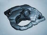 Three-dimensional computed tomography scan of C1.
 The annular fibers are made up of collagenous lamellae with alternating directions of inclination oriented 35° from the horizontal. The annulus is more susceptible to injury with rotation and translation movements due to resistance offered only by the lamella oriented in the direction of movement. In the cervical spine, as in the lumbar spine, the intervertebral disc dissipates the transmission of compressive loads throughout the ROM by slowing the rate at which these forces are transmitted through the spine. By diverting the load via temporarily stretching the annular fibers, the disc protects the vertebra from taking the entire load at once.
In asymmetric loading, the nucleus pulposus migrates toward the area with less load. Thus, in flexion movements of the cervical spine, anterior offset loading of the intervertebral disc occurs, in which the nucleus pulposus moves posteriorly and the posterior annular wall is stretched. In addition, the cervical lordosis reduces, the vertebral canal lengthens, and the intervertebral foramina open.[2]
In extension movements of the cervical spine, posterior offset loading of the intervertebral disc occurs, in which the nucleus moves anteriorly and the anterior annular wall is stretched. Shortening of the vertebral canal and closing of the intervertebral foramen also occur.[2] In lateral flexion and rotation (coupling movement) of the cervical spine, there is offset loading of the intervertebral disc on the side of flexion and rotation, with nuclear material moving to the opposite side (site of the convexity), and the posterolateral annular wall is stretched.[2]
The intervertebral foramina house the exiting cervical nerves. The largest cervical spine foramen is at the C2-3 level, and the smallest foramen is at the C6-7 level.[17] The cervical foramina become very dynamic during cervical spine ROM. The intervertebral foramina enlarge with flexion and decrease with extension. In rotation, the ipsilateral side becomes smaller, and the contralateral side enlarges. The extreme changes of the foramina occur with coupled movements (ie, flexion-rotation and extension-rotation-lateral flexion).[18]
In addition to the above biomechanical concerns, cervical spinal stenosis has been evaluated with regard to catastrophic cervical sports injuries. The Torg/Pavlov ratio (measured by dividing the sagittal diameter of the spinal canal by the sagittal diameter of the vertebral body) when less than 0.8 was thought to subject the football player to high risk of cervical cord injury due to suspected cervical stenosis (see image below). However, subsequent studies found that this ratio may be erroneously low in players that have wide vertebral bodies. A study by Cantu suggested that functional stenosis as documented by myelogram or magnetic resonance imaging (MRI) may be a more appropriate measure of stenosis.[6]
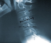 Lateral cervical spine plain radiograph illustrating the Torg/Pavlov ratio. Classification of athletic cervical spine injuries
A review by Bailes and Maroon classified athletes with cervical injuries into 3 types[4] :
Type I injuries were those that caused permanent spinal cord damage, including conditions such as anterior cord syndrome, Brown-Sequard syndrome, central cord syndrome, and mixed incomplete syndrome.Type II injuries were classified as those that occur transiently after athletic trauma with normal neurologic examination and normal radiologic evaluation. Type II injuries included spinal concussion neurapraxia, and "burning hands" syndrome. The burning hands syndrome was described as suspected injury to the spinothalamic and corticospinal tracts, resulting in arm and hand weakness with burning dysesthesias.[19] This is distinct from the burner or stinger injury that is a common cervical injury in football players and is thought to be due to traction on the upper trunk of the brachial plexus. In this condition, athletes typically have a burning, dysesthetic pain that begins in the shoulder region and radiates unilaterally into the arm and hand, with C5-C6 distribution numbness or weakness. Type III injuries were classified in athletes with only radiologic abnormalities but without neurologic deficit. These included congenital spinal stenosis, acquired spinal stenosis, herniated cervical disc, an unstable fracture, fracture/dislocation, ligamentous injury, and spear-tackler’s spine. Spear tackler’s spine was described by Torg et al described athletes that were at high risk for quadriplegic injury. These athletes had developmental cervical canal stenosis, reversal of the cervical lordosis, preexisting posttraumatic cervical radiographic abnormalities, and documentation of using spear-tackling techniques. PreviousProceed to Clinical Presentation , Cervical Discogenic Pain Syndrome


0 comments:
Post a Comment
Note: Only a member of this blog may post a comment.