Background
Medial collateral ligament (MCL) injuries of the knee are very common sports-related injuries. The MCL is the most commonly injured knee ligament. Injuries to the MCL occur in almost all sports and in all age groups.
NextEpidemiologyFrequencyUnited StatesThe incidence of MCL injuries is impossible to determine because of the wide spectrum of injury severity. Many MCL injuries are minor and may never be evaluated by a physician.[1]
PreviousNextFunctional AnatomyThe medial aspect of the knee has been divided into 3 distinct layers based on cadaver dissection. The first layer is the deep fascia, which consists of the sartorius fascia anteriorly and a thin fascial layer posteriorly. The thin posterior fascia covers the popliteal fossa and the heads of the gastrocnemius muscle. The second layer includes the superficial MCL, also known as the tibial collateral ligament. This ligament attaches proximally to the medial femoral epicondyle and to the tibia distally, approximately 4-5 cm distal to the joint line. The parapatellar retinaculum and patellofemoral ligament are within this layer.
The third layer is the knee joint capsule, which attaches proximally and distally at the articular margins. The capsule is divided into thirds from anterior to posterior. The anterior third of the capsule is the thinnest portion. It is attached to the anterior horn of the medial meniscus and is reinforced by the medial retinaculum. The middle third of the capsule consists of the deep medial collateral ligament. It is firmly attached to the mid body of the medial meniscus. Proximal to the meniscal attachment, it is termed the meniscofemoral ligament. Distal to its meniscal attachment, it is termed the meniscotibial ligament. The posterior third of the capsule includes the posterior oblique ligament (POL) and the oblique popliteal ligament. The POL has 3 arms, the superficial, tibial, and capsular.
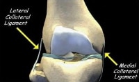
See the figure below.
 The medial and lateral collateral ligaments of the knee. Courtesy of Randale Sechrest, MD, CEO, Medical Multimedia Group PreviousNextSport Specific Biomechanics
The superficial MCL has been shown through serial cutting studies to provide the primary restraint to valgus loads at all degrees of flexion. It is also an important restraint to anterior tibial translation when the anterior cruciate ligament is injured. The superficial MCL acts as a primary restraint to external rotation of the tibia.
Stability of the medial side of the knee is provided by dynamic and static restraints. The static restraints are the superficial MCL and the joint capsule, including the deep MCL and the POL. The semimembranosus muscle, the pes anserine muscles, and the vastus medialis muscle provide dynamic stability. The muscles of the pes include the sartorius, gracilis, and semitendinosus. These muscles flex and internally rotate the tibia. The semimembranosus has 4 attachments: direct, tibial, inferior, and capsular.[2, 3]
PreviousProceed to Clinical Presentation , Medial Collateral Knee Ligament Injury


0 comments:
Post a Comment
Note: Only a member of this blog may post a comment.