Background
Although hamate fractures are increasing in incidence secondary to the popularity of sports activities involving racquets, bats, and clubs, these injuries remain relatively rare. Estimates suggest hamate fractures constitute 2% of all carpal fractures. The hamate bone is a roughly triangular-shaped bone composed of both a body and a hook (see images below). Hamate fractures are thus classified as type I fractures involving the hook and type II fractures involving the body. Type I fractures are more common than type II fractures.[1, 2]
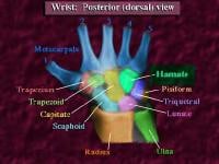 Posterior (dorsal) view of the wrist.
 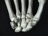 Anterior palmar view.
 For excellent patient education resources, visit eMedicineHealth's First Aid and Injuries Center. Also, see eMedicineHealth's patient education articles Wrist Injury and Broken Hand.
Related Medscape Reference topics:
Carpal Fractures
Wrist Fracture in Emergency Medicine
Wrist Fractures and Dislocations
Metacarpal Fractures
Related Medscape resources:
Resource Center Exercise and Sports Medicine
CMEÂ Physical Exercise May Help Reduce Fatigue During and After Cancer Treatment
CMEÂ Regular Exercise Through Middle Age May Delay Biological Aging
CME/CEÂ Risks and Benefits of Exercise Reviewed in AHA Statement
NextEpidemiologyFrequencyUnited StatesHamate fractures account for 2% of all carpal fractures. Of the 2%, one third are hamate hook fractures due to repetitive swinging by golfers.
PreviousNextFunctional AnatomyThe hamate is a triangular bone located in the distal carpal row farthest to the ulnar side (see the images below). The hamate is bordered proximally by the pisiform and the lunate in the proximal carpal row, radially by the capitate, and distally by the bases of the fourth and fifth metacarpals.
Posterior (dorsal) view of the wrist. Anterior palmar view. A roughly circular projection or hook on the volar surface of the hamate is the inferolateral border of the Guyon canal. The roof (superficial) of the canal is formed by the palmar carpal ligament, and the floor (deep) is formed by the flexor retinaculum. The canal carries the ulnar artery and nerve, and, for this reason, hook fractures should suggest a high probability of ulnar artery and nerve damage.[3] In addition, the hamate hook has a dual blood supply, with vessels entering from both the ulnar tip and radial base. These vessels often have a poor anastomosis, which clinically can result in nonunion due to insufficient blood supply.
PreviousNextSport-Specific BiomechanicsType I fractures involving the hook of the hamate are the most common and can occur via several different mechanisms.[1, 2, 4, 5, 6, 7] First, repeated microtrauma to the hook during sports involving swinging clubs, bats, or racquets can result in a hook stress fracture. These usually occur in the nondominant hand and account for approximately one third of hamate fractures. Second, direct trauma can be applied during sports when the butt of the club rests on the hamate and the force of the swing is then transmitted directly to the bone. In addition, indirect trauma can be applied to the hook through its muscular and ligamentous attachments. This can occur either when falling on a hyperextended wrist or during power grips.
Type II fractures involving the body of the hamate are less common than type I fractures and always require direct force.[4] Most commonly, these fractures occur with a punch-press injury or dorsopalmar compression of the wrist between heavy weights.
Related Medscape resource:
Resource Center Exercise and Sports Medicine
PreviousProceed to Clinical Presentation , Hamate Fracture


0 comments:
Post a Comment
Note: Only a member of this blog may post a comment.