Background
Elbow dislocation is the most common dislocation in children; in adults, it is the second most common dislocation after that of the shoulder.[1, 2, 3, 4, 5] The elbow is amazingly stable, relying more on bony anatomy configuration for stability rather than ligaments. Considerable force is necessary to dislocate the elbow; sports activities account for up to 50% of elbow dislocations, and this type of injury is more commonly seen in adolescent and young adult populations (see the image below).
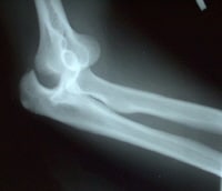 Posterior and lateral dislocation of the left elbow in a soccer goalie. A small avulsion fracture of the olecranon is present.
 Posterior elbow dislocations comprise over 90% of elbow injuries. Early recognition of this injury is required due to the need for early reduction, given a higher likelihood for poor function and possible neurovascular compromise with delays in reduction.[1, 2, 3, 4, 6, 7] Associated fractures are not infrequent with elbow dislocations, given the force that is required to dislocate the elbow.
Anterior dislocations are seen much less commonly than posterior dislocations. Divergent dislocations, which result in the ulna and radius dislocating in opposite directions, are even more rare. In the pediatric population, radial head subluxation is the main cause of elbow dislocations.
For excellent patient education resources, visit eMedicineHealth's First Aid and Injuries Center. Also, see eMedicineHealth's patient education articles Elbow Dislocation and Broken Elbow.
NextEpidemiologyFrequencyUnited StatesThe rate of elbow dislocation is 6-13 cases per 100,000 people, and this injury occurs more frequently in males than in females. Of all elbow dislocations, 10-50% are sports related. More than 90% of elbow dislocations are posterior dislocations.
PreviousNextFunctional AnatomyThe elbow is primarily a flexion-extension hinge joint, which also allows for pronation and supination. Normal range of motion (ROM) at the elbow should be extension to 0° and flexion to 150°.[1, 2]
The humerus and ulna form a very stable unit, which is generally resistant to disruption unless considerable force is applied. This inherent stability also reduces the likelihood of redislocation. The primary bony stabilizers are the coronoid and radial head.
The medial collateral ligament (MCL) and lateral collateral ligament (LCL) comprise the ligamentous stability of the elbow and act as a back-up system to the elbow's natural bony stability. The MCL consists of 3 bands, the anterior oblique, posterior oblique, and the transverse. The anterior band provides most of the resistance to valgus stress. The LCL has 2 bands, the ulnar collateral and radial collateral.
The 2 main compartments of the elbow are the anterior and posterior compartments. The anterior compartment contains the brachial artery and the ulnar and median nerves. This compartment is more commonly affected by dislocations and is the reason for clinical concern regarding brachial artery disruption and median or ulnar nerve entrapment.[1, 2, 4, 8]
The ulnar nerve passes posteriorly to the medial epicondyle of the humerus, and then it travels deep in the forearm before becoming more superficial again at the wrist. The close proximity of the ulnar nerve to the medial epicondyle allows for the increased likelihood of entrapment when a dislocation occurs. The median nerve is also frequently affected and travels intimately with the brachial artery, which predisposes to simultaneous injury for both the artery and nerve. The posterior compartment contains the radial nerve and triceps brachii muscle.
Anatomically, the mechanism for elbow dislocations is believed to occur as a continuum of damaged/torn structures, beginning laterally with the ulnar portion of the LCL, followed by complete LCL disruption, then damage to the anterior and posterior compartments. The posterior MCL can then become damaged, leaving the anterior portion intact. Further force can allow the elbow to pivot about the anterior bundle of the MCL, potentially damaging it. The LCL, therefore, is considered to be the initial weak link in elbow dislocations.
In the pediatric population, the clinician should be aware of the 6 ossification centers of the elbow joint as well as the annular ligament. The capitellum, radial head, internal (medial) epicondyle, trochlea, olecranon, and external (lateral) epicondyle (CRITOE) is the order in which the ossification centers appear. These centers may often be mistaken for fractures on x-rays. NOTE: A general rule of thumb for the time of appearance of the ossification centers is "1-3-5-7-9-11," which are the ages in years, corresponding to the CRITOE pneumonic.
In cases in which there is radial head subluxation, the radial head slips under the annular ligament and becomes trapped.
PreviousNextSport-Specific BiomechanicsBiomechanically, no single sport definitively increases the risk of elbow dislocations; however, sports that increase the likelihood of a person falling onto an outstretched hand (ie, FOOSH injury) (eg, gymnastics, rollerblading, cycling) may theoretically increase the risk of elbow dislocation.
PreviousProceed to Clinical Presentation , Elbow Dislocation


0 comments:
Post a Comment
Note: Only a member of this blog may post a comment.