Background
For as much as we use our hands, it is surprising that they are not injured more frequently. Sports-related metacarpal fractures most commonly occur during participation in contact sports, such as football, rugby, or basketball, in which the hands are unprotected. A direct fall onto the hand (FOOSH injury) while cycling, running, or skiing may also result in a fracture.
See the images below.
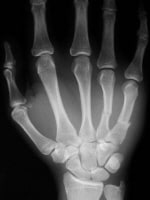 Displaced fourth and fifth metacarpal fractures, anteroposterior view.
 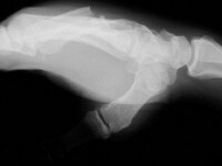 Displaced fourth and fifth metacarpal fractures, lateral view.
 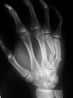 Fourth and fifth metacarpal fractures, oblique view.
 For patient education resources, see the Fractures and Broken Bones Center, as well as Boxer's Fracture, Broken Hand, Cast Care, Finger Dislocation, Finger Injuries, and Human Bites.
Related Medscape Reference topics:
Fracture, Hand
Hand, Fracture and Dislocations: Metacarpal [in the Plastic Surgery section]
Hand, Fracture and Dislocations: Phalangeal
Metacarpal Fractures [in the Orthopedic Surgery section]
Related Medscape topics:
Resource Center Adolescent Medicine
Resource Center Exercise and Sports Medicine
Specialty Site Orthopaedics
Conservative Treatment for Closed Fifth (Small Finger) Metacarpal Neck Fractures
Until the early 20th century, metacarpal fractures were treated exclusively by nonoperative means. Surgery was first suggested as an alternative treatment for certain fracture patterns in the 1920s.[1, 2]
NextEpidemiologyFrequencyUnited StatesMetacarpal and phalangeal fractures are the most common fractures of the upper extremity. They account for approximately 10% of all orthopedic fractures.[2, 3] Most occur in young adults, usually as a result of direct blunt trauma, axial loading, or throwing a punch during an altercation. The thumb and small finger are the most frequently injured.
PreviousNextFunctional AnatomyThe finger metacarpals describe a gentle arch in both the axial and coronal planes. Each bone is relatively straight along its dorsal cortex and concave along the palmar surface.
The carpometacarpal (CMC) joints consist of 5 metacarpal bases that articulate with the trapezoid, trapezium, capitate, and hamate. Articular congruity of the joint surfaces, in combination with the strong interosseous and extrinsic palmar and dorsal ligaments, provides stability to the CMC joint. The CMC joints of the index and long fingers are essentially fixed, whereas those of the ring and small fingers enjoy 20-30° of motion in flexion-extension. The thumb is extremely mobile at the CMC joint.
The opposing saddle shapes of the metacarpal base and the articulating trapezium allow for flexion, extension, abduction, and adduction. The joint capsule and ligaments permit a small degree of rotation. The most important soft-tissue support for the first CMC joint is the anterior oblique ligament, which runs from the tubercle of the trapezium to the volar beak of the metacarpal. This ligament may be ruptured in a dislocation, but it is most commonly avulsed by a fragment of bone from the ulnar corner of the metacarpal (Bennett fracture).[4]
PreviousNextSport-Specific BiomechanicsThe anatomic relationships described above maintain proper rotational alignment of the fingers and allow for the smooth production of power grip and the ability to clench the fist, functions that are required in many sports. The high mobility of the thumb enables both pinching (squeezing small equipment or objects between the thumb and the forefinger) and grasping of large objects.
Bennett fractures are unstable because of the deforming forces of the intrinsic and extrinsic muscles. The anterior oblique ligament stabilizes the volar-ulnar fragment, but the thenar muscles and abductor pollicis longus displace the remaining metacarpal in the proximal, dorsal, and radial directions.[5]
An analogous situation exists with the reverse Bennett fracture of the small-finger metacarpal. Intermetacarpal ligaments stabilize the radial fragment. The hypothenar and the flexor and extensor carpi ulnaris muscles pull the remaining metacarpal proximally and dorsally.
PreviousProceed to Clinical Presentation , Metacarpal Fracture and Dislocation


0 comments:
Post a Comment
Note: Only a member of this blog may post a comment.