Background
With an increase in public interest in physical fitness, clinical practitioners are diagnosing stress fractures with greater frequency.[1, 2, 3] First described by Aristotle in 200 BC, stress fractures were initially recorded in the medical literature in 1855 by the Prussian military physician Breithaupt, who described what is now known as a march fracture, or stress fracture of the metatarsals. See the images below.
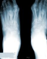 Radiograph of the feet. This image depicts a stress fracture of the left second metatarsal with exuberant callus.
  Radiograph of the left foot. This image depicts a stress fracture of the fifth metatarsal.
 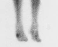 Bone scan of the lower extremities. This image depicts a right fifth metatarsal stress fracture.
 Metatarsal stress fractures are not limited to high-level athletes or military recruits. This type of injury is seen in runners of all levels, as well as ballet dancers and gymnasts and patients with rheumatoid arthritis (RA), metabolic bone disease, and neuropathic conditions.[4, 5, 6] Metatarsal stress fractures are also seen with increasing frequency in patients who engage in aerobics activities, particularly high-impact aerobics.
NextEpidemiologyFrequencyUnited StatesThe incidence of stress fractures in the general population is unknown, as virtually all literature on the subject is derived from a military population or advanced-level athletes. Stress fractures are estimated to constitute up to 16% of all injuries that are related to athletic participation; running is the cause in most of these cases. Most stress fractures (95%) involve the lower extremities, particularly the metatarsals.
PreviousNextFunctional AnatomyThe second and third metatarsals are relatively fixed in position within the foot; the first, fourth, and fifth metatarsals are relatively mobile. More stress is placed on the second and third metatarsals during ambulation; thus, these bones are at increased risk for stress fractures.
The fifth metatarsal, which is approximately 1.5 cm from the proximal pole of the bone, bears greater stress in those who oversupinate when they walk or run. The fifth metatarsal also has a diminished blood supply and, thus, a decreased ability to heal.[7, 8]
Stress fractures of the proximal fifth metatarsal must be distinguished from proximal avulsion fractures ("pseudo-Jones" fractures) and Jones fractures. The proximal avulsion fracture is usually associated with a lateral ankle strain and occurs at the insertion of the peroneus brevis tendon. The true Jones fracture is an acute fracture of the proximal diametaphyseal junction.
PreviousNextSport-Specific BiomechanicsQueen et al investigated whether foot type (flat or normal) resulted in loading differences during four sport-specific tasks (cross-cut, side-cut, shuttle run, and landing from a simulated lay-up).[9] Of 22 healthy individuals, 12 had normal feet and 10 had flat feet, and each completed 5 trials per condition. In-shoe pressure data were collected at 50 Hz, and analyses of the entire foot and 8 regions of the foot were carried out on contact area, maximum force, and the force time integral. The investigators' findings included the following statistically significant (P [9] :
Flat feetDuring the cross-cut task, there was an increase in medial midfoot contact area.During the side-cut task, an increase in contact area, force time integral, and maximum force in both the medial and lateral midfoot were demonstrated. During the shuttle run task, an increase in force time integral in the lateral midfoot and increases in maximum force in both the medial and lateral midfoot were present During the landing task, an increase in maximum force in the medial midfoot was present. However, flat feet showed a decrease in middle forefoot maximum force.Queen et al concluded that individuals with a normal foot may have a lower risk for medial and lateral midfoot injuries such as metatarsal stress fractures. Thus, foot type should be assessed when determining an individual's risk for metatarsal stress fractures.[9]
PreviousProceed to Clinical Presentation , Metatarsal Stress Fracture


0 comments:
Post a Comment
Note: Only a member of this blog may post a comment.