Background
Injuries in and around the shoulder are common in today's athletic society. Proper knowledge of the different problems and treatment options for shoulder disorders is necessary to get patients back to their preinjury state.
Acromioclavicular (AC) joint injuries are common and often seen after bicycle wrecks, contact sports, and car accidents. The acromioclavicular joint is located at the top of the shoulder where the acromion process and the clavicle meet to form a joint. Several ligaments surround this joint, and depending on the severity of the injury, a person may tear one or all of the ligaments. Torn ligaments lead to acromioclavicular joint sprains and separations.[1]
The distal clavicle and acromion process can also be fractured. Injury to the acromioclavicular joint may injure the cartilage within the joint and can later cause arthritis of the acromioclavicular joint.
This article discusses the anatomy of the acromioclavicular joint, the diagnosis of disorders of this joint, and the different treatment options.
For excellent patient education resources, see eMedicineHealth's First Aid & Injuries Center. Also, see eMedicineHealth's article on Shoulder Dislocation.
NextEpidemiologyFrequencyUnited StatesInjuries to the acromioclavicular joint are the most common reason that athletes seek medical attention following an acute shoulder injury. Glenohumeral dislocations (see Shoulder Dislocation) are the second most common injuries seen. Men in their second through fourth decades of life have the greatest frequency of acromioclavicular joint injuries, which are most often incomplete tears of the ligaments.[1]
PreviousNextFunctional AnatomyThe normal width of the acromioclavicula joint is 1-3 mm in younger individuals; it narrows to 0.5 mm or less in individuals older than 60 years.
The acromioclavicular joint is made up of 2 bones (the clavicle and the acromion), 4 ligaments, and a meniscus inside the joint.
The acromioclavicular joint is surrounded by a thin joint capsule and 4 small ligaments. These ligaments mostly give joint stability to anterior and posterior translation, as well as provide horizontal stability to the joint.
Another set of ligaments also provides vertical stability to the acromioclavicular joint. These ligaments are called the coracoclavicular ligaments, which are found medial to the acromioclavicular joint and go from the coracoid process on the scapula to the clavicle.
Different injuries result in different tears of the 2 coracoclavicular ligaments (the conoid and the trapezoid). Torn acromioclavicular joint ligaments and/or torn coracoclavicular ligaments are seen in acromioclavicular joint sprains. The meniscus that lies in the joint may also be injured during sprains or fractures around the acromioclavicular joint. The acromioclavicular capsular ligaments provide most of the joint stability in the anteroposterior (AP) direction. The conoid and trapezoid ligaments aid in providing superior-inferior stability to the joint. Compression of the joint is restrained mainly by the trapezoid ligament.
PreviousNextSport-Specific BiomechanicsWhen a person falls onto their shoulder, the force pushes the tip of the shoulder down. The clavicle is usually kept in its anatomic position, whereas the shoulder is driven down, which injures the different ligaments or causes a fracture. When the ligaments are injured they are either sprained or, in more severe cases, torn.
Acromioclavicular joint sprains have been classified according to their severity. In a type I sprain, a mild force applied to these ligaments does not tear them. The injury simply results in a sprain, which hurts, but the shoulder does not show any gross evidence of an acromioclavicular joint dislocation. Type II sprains are seen when a heavier force is applied to the shoulder, disrupting the acromioclavicular ligaments but leaving the coracoclavicular ligaments intact. When these injuries occur, the lateral clavicle becomes a little more prominent.
In type III sprains, the force completely disrupts the acromioclavicular and coracoclavicular ligaments. This leads to complete separation of the clavicle and obvious changes in appearance. The lateral clavicle is very prominent. A few more types of acromioclavicular joint sprains have been classified, but types I–III are the most common (see below).
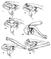 Classification of acromioclavicular joint injuries.
 An acromioclavicular joint sprain is more common than a fracture after an injury. However, fractures of the distal clavicle and the acromion process may occur, so the healthcare provider must be aware of such injuries and ready to diagnose and treat them as well (see Clavicular Injuries).
PreviousProceed to Clinical Presentation , Acromioclavicular Joint Injury


0 comments:
Post a Comment
Note: Only a member of this blog may post a comment.