Friday, January 31, 2014

Little League Elbow Syndrome

Background

Little league elbow (LLE) syndrome is a valgus overload or overstress injury to the medial elbow that occurs as a result of repetitive throwing motions. Over the past several decades, the number of organized sports for children has grown significantly, with millions of children participating in organized athletics each year. This increase in participation has been paralleled by an increase in sports-related injuries in the pediatric population.[1, 2, 3, 4, 5, 6, 7, 8, 9, 10, 11]

Increased single-sport participation with year-round training, higher intensities at young ages, and longer competitive seasons are contributing factors to the increased injury rates seen in pediatric athletes. Conditioning and training errors also contribute significantly to the risk and frequency of injury. Although briefly discussed below, injuries to the lateral, posterior, and anterior elbow are separate entities and should not be confused with the medial injuries referred to as little league elbow syndrome.

During the throwing motion, valgus stress is placed on the elbow. This valgus stress results in tension on the medial structures (ie, medial epicondyle, medial epicondylar apophysis, medial collateral ligament complex) and compression of the lateral structures (ie, radial head, capitellum). Repeated stress results in overuse injury when tissue breakdown exceeds tissue repair. Recurrent microtrauma of the elbow joint can lead to little league elbow, a syndrome that encompasses (1) delayed or accelerated growth of the medial epicondyle (medial epicondylar apophysitis), (2) traction apophysitis (medial epicondylar fragmentation), and (3) medial epicondylitis.[1, 8, 12, 13, 14, 15]

Medial epicondylar apophysitis and stress fractures through the medial epicondylar epiphyses caused by repetitive valgus stress generally manifest with progressive medial pain, decreased throwing effectiveness, and decreased throwing distance.

Other causes of medial elbow pain include avulsion fractures of the medial epicondyle and ulnar collateral ligament (UCL) sprains or tears. Although a fracture is usually an acute traumatic event, a preceding history of medial elbow pain is common and is thought to be a risk factor for progression to acute fracture. Therefore, any thrower who is experiencing medial elbow pain should refrain from pitching until he or she has had a thorough evaluation.[1, 3, 5, 8, 16]

A medial epicondyle fracture manifests as point tenderness and swelling over the medial epicondyle, often with an elbow flexion contracture greater than 15°. Repetitive medial stress can also cause attenuation and microstretching of the UCL complex, causing mild instability over time.[1]

UCL injuries can manifest as acute ligament tears following a single valgus stress or as overuse sprains following repetitive valgus overloads. The clinical presentation is similar to little league elbow; however, the typical age range of the athlete is the older teenager who is skeletally mature. Suspected UCL injuries should be referred for further evaluation by a sports medicine specialist. Athletes with UCL injuries should not be allowed to pitch until they have been evaluated.

Although uncommon in children, neurologic injuries such as C8-T1 radiculopathy and ulnar neuritis can manifest as medial elbow pain and should be included in the differential diagnosis (see Differentials and Other Problems to Be Considered).

Lateral compression of the elbow most frequently results in injuries to the capitellum and radial head. Osteochondrosis of the capitellum (known as Panner disease) generally occurs in children aged 7-12 years and manifests as dull, achy, activity-related lateral elbow pain. Swelling, clicking, and decreased range of motion are uncommon associated symptoms. Panner disease tends to be a benign self-limited condition that does well over time and is treated with complete rest from inciting activities such as throwing and weight bearing on the elbow. Osteochondral injuries can also be observed in the radial head.

Osteochondritis dissecans (OCD) of the capitellum occurs in adolescents aged 13-17 years. This is a localized injury to subchondral bone that results from repetitive lateral compression of the elbow during overhead motions. These patients report a general dull elbow pain that worsens with activity, often have a flexion contracture of 15° or greater, and may have mechanical symptoms of clicking or popping. Loose body formation, residual capitellum deformity, and elbow degenerative joint disease are potential sequelae. Different treatment options are used based on the age and skeletal maturity of the patient and the type of lesion present.

Osteochondritis dissecans lesions can be separated into type I, which has no displacement and no articular cartilage fracture; type II, which has evidence of articular cartilage fracture or partial displacement; and type III, which is completely displaced with loose bodies in the joint.

Posterior elbow injuries also occur as a result of throwing. During the follow-through stage of throwing, extension overload and valgus stress can result in injury of the olecranon. These athletes present with posterior elbow pain, clicking, and possible loss of elbow extension. Loose bodies and olecranon nonunion can occur in younger athletes. Older athletes may experience olecranon fractures or secondary osteophyte formation. These injuries are sometimes treated surgically.[1, 3, 4, 5, 6, 7, 9, 10, 11, 17]

For excellent patient education resources, visit eMedicineHealth's First Aid and Injuries Center. Also, see eMedicineHealth's patient education article Repetitive Motion Injuries.

NextEpidemiologyFrequencyUnited States

Annually, an estimated 4.8 million children aged 5-14 years participate in baseball and softball. The incidence of all baseball-related overuse injuries is 2-8% per year. The incidence of overuse injuries in the 9- to 12-year-old range for baseball is 20-40%, and in the adolescent age group is 30-50%. The true incidence of sports-related injuries is unknown because a large number of athletes never seek medical care. Early recognition of little league elbow syndrome is important, because it leads to better outcomes and decreases the risk of persistent functional disabilities in the athletes.

International

No data are available for the annual incidence of little league elbow syndrome in the international community.

PreviousNextFunctional Anatomy

Evaluation of the young adolescent elbow presents some anatomic challenges to the healthcare provider in that the elbow consists of numerous ossification centers and cartilaginous physes. Becoming familiar with the chronologic order of appearance and ossification of these growth centers is important. Consider the mnemonic CRITOE (ie, capitellum, radius, internal epicondyle, trochlea, olecranon, external epicondyle).

Each of the ossification centers appears at a relatively predictable time starting around age 1-2 years, with 2-year intervals between the next center's appearance. Closure of each of the apophyses occurs from age 14 to 16 years, with the medial epicondyle specifically closing at approximately age 15 years. The elbow likely reaches full skeletal maturity by the late teen years, at which time injuries to the UCL are far more common. Until then, the young thrower is at risk for little league elbow syndrome.[1, 4, 12, 16, 18]

The static stabilizers around the elbow include the bony articulations, the joint capsule, and the various ligament bundles. The medial (ulnar) collateral ligamentous complex consists of the anterior oblique bundle, posterior oblique bundle, and transverse ligament. These structures are the primary medial support of the elbow during valgus stress. The lateral (radial) ligamentous complex, composed of the lateral collateral, lateral ulnar collateral, and accessory lateral collateral ligaments, provides support during varus stress.

The dynamic stabilizers primarily include the muscles that cross the elbow joint, such as the triceps, biceps, and brachioradialis. The flexor-pronator group stabilizes against valgus stress, and the extensor-supinator group stabilizes against varus stress.

Elbow biomechanics include flexion/extension range of motion and pronation/supination. Slight hyperextension 5-15° through flexion of approximately 150° is within normal limits. Baseball pitchers with years of throwing experience often have relative 5-10° flexion contractures on their dominant side; however, in the young thrower, a flexion contracture can be a sign of injury. Pronation of 75° and supination of 85° is normal. Varus-valgus laxity of 3-4° is normal.

PreviousNextSport-Specific Biomechanics

One should be familiar with the stages of throwing to understand the complexities of the biomechanical forces that contribute to the young thrower's risk of injury, such as in little league elbow syndrome. The pitching or throwing motion can be divided into 6 stages. Medial elbow injuries are the most common type seen in throwers and occur most commonly in the cocking and acceleration phases of throwing, owing to the presence of maximum valgus extension or distraction forces.[1, 12, 13, 14, 15]

Windup begins with the pitcher balancing his weight over his rear leg, with the elbow flexed and the forward leg flexed at least 90°. Stride starts with the lead leg beginning to descend toward the plate, and the 2 arms separate. The elbow moves from extension into flexion of 80-100°. Cocking occurs when the humerus is in extreme abduction and external rotation and the elbow is flexed. The lead foots contacts the ground, the pelvis and trunk rotate, and elbow torque transfers valgus force across the elbow joint. During this phase, medial tension and lateral compression forces are applied to the elbow. Acceleration is the shortest pitching phase, lasting from maximal external shoulder rotation to ball release. In this phase, the trunk rotates as the elbow extends. Maximum elbow angular velocity is comparable during fastballs, sliders, and curveballs, but it less during the change-up pitch. Velocity comes from rotation of the trunk, shoulder, and hips. Varus torque forces during this phase act to resist the valgus extension "overload" phenomenon and can contribute to posterior elbow (olecranon) impingement. Deceleration is initiated at ball release and ends when the shoulder has reached full internal rotation. The body must decelerate the arm and dissipate forces in the elbow and shoulder. Follow-through is the final phase of the baseball pitch and ends with the pitcher reaching a balanced fielding position with full-trunk rotation and the body weight fully transferred from the rear leg to the forward leg. During follow-through, the elbow flexes into a relaxed position and crosses the body. PreviousProceed to Clinical Presentation , Little League Elbow Syndrome

Humeral Capitellum Osteochondritis Dissecans

Background

In 1889, Francis Konig described osteochondritis dissecans as a subchondral inflammatory process of the knee resulting in a loose fragment of cartilage from the femoral condyle. Although no inflammatory cells have been identified on histologic sections of excised fragments, the term osteochondritis dissecans has persisted and since been broadened to describe a similar process occurring in many other joints, including the knee, hip, ankle, elbow, and metatarsophalangeal joints.[1, 2, 3, 4, 5, 6]

Humeral capitellum osteochondritis dissecans occurs after the capitellum has ossified and is the result of "injury" to the subchondral bone. The initial histologic appearance is consistent with avascular necrosis. The avascular necrosis of subchondral bone leads to loss of support for adjacent cartilaginous structures. The natural history of some osteochondritis dissecans lesions is the separation of these structures from the capitellum, leading to the development of an osteochondral fragment of articular cartilage on the underlying bone at the superficial surface of the diarthrodial joint.[7, 8, 9, 10]

For excellent patient education resources, see eMedicineHealth's patient education article Tennis Elbow.

Related Medscape Reference topics:

Elbow and Forearm Overuse Injuries

Lateral Epicondylitis

Medial Epicondylitis

Overuse Injury

Related Medscape resources:

Resource CenterExercise and Sports Medicine

Specialty SiteOrthopaedics

Specialty SitePathology & Lab Medicine

American Orthopaedic Society for Sports Medicine 31st Annual Meeting-Competing Tennis Elbow Surgeries Both Deemed Successful

Deep transverse friction massage for treating tendinitis

Physical Therapy and Brace Therapy for Tennis Elbow

NextEpidemiologyFrequencyUnited States

Humeral capitellum osteochondritis dissecans comprises 6% of all osteochondritis dissecans cases.

In the United States, humeral capitellum osteochondritis dissecans most commonly occurs in the second decade of life and is rare in individuals younger than 10 years or older than 50 years. Humeral capitellum osteochondritis dissecans is primarily observed in children aged 10-15 years.

Approximately 85% of osteochondritis dissecans cases involve males, with a large proportion of these being Little League pitchers. Humeral capitellum osteochondritis dissecans is believed to affect 4.1 of every 1000 males. Among male relatives of affected males, the prevalence rate is 14.6%. Osteochondritis dissecans also occurs in females, most notably gymnasts. Finally, it also commonly occurs in persons who participate in racquet sports and in weight lifting.

Humeral capitellum osteochondritis dissecans usually occurs in the dominant arm. In up to 20% of cases, it occurs bilaterally.

PreviousNextFunctional Anatomy

While the trochlea of the distal humerus articulates with the sigmoid fossa of the proximal ulna, the capitellum of the distal humerus articulates with the head of the radius. These articulations, in conjunction with the radioulnar articulation, compose the elbow joint. The articulation of the radial head and humeral capitellum provides mobility for a wide range of supination and pronation, as well as flexion and extension. This area is thus particularly susceptible to the rotary, compressive, axial, and angular forces associated with activities such as throwing.

The radiocapitellar articulation is supported laterally by the radiocollateral, the accessory collateral, the lateral ulnar collateral, and the annular ligaments. These ligaments function to stabilize the elbow throughout the motions of pronation, supination, flexion, and extension.

PreviousNextSport-Specific Biomechanics

The exact etiology of osteochondritis dissecans is unclear.[1, 4, 5, 11, 12, 13] In overhead throwing, articular forces at the radiocapitellar articulation are significant. Progressive pronation, compression, and rotation occur on the anteromedial radial head and the inferior and medial aspects of the capitellum as the elbow is extended.

These forces are believed to lead to fibrillation on the articular surface and subchondral osseous changes, with the possible production of osteocartilaginous fragments and the development of humeral capitellum osteochondritis dissecans. The valgus orientation of the elbow contributes to these compressive loads. Excessive axial loading to the elbow is also believed to be the primary cause of injury in gymnasts and weight lifters.[14, 15]

PreviousProceed to Clinical Presentation , Humeral Capitellum Osteochondritis Dissecans

Thursday, January 30, 2014

Gamekeeper's Thumb

, Gamekeeper's Thumb

Wednesday, January 29, 2014

Hand Dislocation

background

Hand dislocation is a common damage in sports activities and in occupational settings, incessantly appearing to be minor. If the athlete, trainer, or show has already reduced the dislocation, it appears unimpressive when put next with an incredible knee damage or a shoulder dislocation.

however, hand dislocations have actual potential for long-time period incapacity in sports activities and other areas of existence if enough discount is not carried out, if associated injuries are not recognized and as it should be handled or referred, and if doable issues of the damage and its therapy aren't foreseen. The judgment of the initial treating medical doctor may also be crucial in determining the lengthy-time period outcome of these accidents.

Many hand dislocations will also be successfully handled with closed discount, traction, or each. Grossly unstable joints and people for which closed discount has failed most often require surgical intervention. physical and occupational therapy are key elements of therapy all through. Any long-term problems (usually involving stiffness or instability) that improve must be addressed.

NextAnatomyInterphalangeal joints

The bony anatomy of the proximal interphalangeal (PIP) joint includes medial and lateral condyles on the proximal phalanx, with matching concavities on the associated distal phalanx. The joint has a variety of motion (ROM) in flexion and extension but is moderately rigid in abduction and adduction; hence, it is a hinge (ginglymus) joint functionally. The bony anatomy of the distal interphalangeal (DIP) joint is an identical, but the surrounding soft tissue gives extra restriction in flexion.

The extrinsic flexors across each joints are as a minimum four occasions greater than the extensors, permitting flexion contractures to strengthen very abruptly, especially with immobilization in flexion. sufficient ROM, particularly at the PIP joint, is critical for normal hand function.

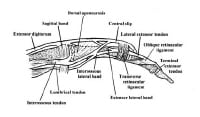
The PIP and DIP joints are both supported on all 4 sides by using an identical tender-tissue buildings, which embody the volar plate on the palmar side (the integrity of which is essential for a secure reduction), collateral ligaments on the radial and ulnar sides, and the extensor complicated (vital slip, lateral bands, and hood) dorsally (see the image under). These buildings connect to and beef up the joint tablet. For a dislocation to occur, at least 1, regularly 2, and sometimes 3 of those constructions have to be considerably injured.

Lateral view of relevant finger anatomy. Lateral view of relevant finger anatomy.

The volar plate is a roughly triangular structure with its base oriented distally, attaching to the volar base of the center phalanx with its tip attaching to the distal facet of the proximal phalanx. The volar plate features generally in limiting hyperextension. to that end, it's just about at all times injured in dorsal dislocations.

The collateral ligaments restrict the joint from opening to varus or valgus stress and are additionally commonly injured in dorsal dislocation. injury to the radial collateral ligament is about 6 occasions more fashionable than harm to the ulnar collateral ligament.

The extensor complex comprises the imperative slip, which attaches to the base of the middle phalanx; the lateral bands, which run dorsolaterally on each and every aspect; and the transverse retinacular ligament, which connects these buildings and extends laterally. It helps restrict volar movement of the base of the center phalanx and subsequently is frequently injured in volar dislocations at the PIP joint, with the center phalanx both tearing the principal slip from its insertion or buttonholing throughout the transverse retinacular ligament between the significant slip and a lateral band.

Metacarpophalangeal joint

The metacarpophalangeal (MCP) joint is regarded as an ellipsoid joint. the head of the metacarpal includes medial and lateral condyles and is narrower on its dorsal floor than on its palmar floor; it fits into the concavity of the bottom of the proximal phalanx. The true collateral ligament attaches to a recess created by the junction of the shoulder and head. The collateral ligament is composed of the next 2 elements:

A dorsally positioned twine portionA fan-formed volar portion or accessory collateral ligament, which extends from the metacarpal to the perimeters of the volar plate

to accomplish flexion and extension on the MCP joint, the anterior and posterior components of the tablet should be lax. When the joint is prolonged, the phalanges have substantial lateral play in abduction and adduction; in consequence, this joint shouldn't be regularly injured. on the other hand, if the ligament is torn, dislocation happens.

The MCP joint of the thumb has radial and ulnar collateral ligaments, which might be loose when the joint is prolonged and tight when flexed. When the joint is extended, the proximal phalanx has the lateral play finished by way of the motion of the interosseous muscles.

When the thumb is flexed and in a practical position, as in the case of many sports activities eventualities (eg, snowboarding, falls on a gloved hand), the ulnar collateral ligament is the structure in danger and will also be ruptured (see Skier’s Thumb). The ulnar collateral ligament can then be displaced in order that the adductor aponeurosis is interposed between the ruptured end of the ligament and its web page of bony attachment.

Carpometacarpal joint

The bony anatomy of the carpometacarpal (CMC) joint includes the 5 metacarpal bases that articulate with the trapezoid, trapezium, capitate, and hamate (in that order) from the radial side of the hand to its ulnar aspect. The CMC joint is a moderately fixed joint section because of the articular congruity of the joint surfaces, with the metacarpal bases acting like concave receptacles to the distal carpal row, and because of the strong interosseous and extrinsic ligament complicated.

The palmar and dorsal ligaments are dissimilar, with the palmar ligaments being better. The scaphoid acts as a link between the proximal and distal carpal rows. The extensor and flexor tendons move over this articular area but add no strength to the CMC joint because the bases of the metacarpals dislocate dorsally relative to the distal carpal row.

the first CMC joint (also referred to as the first metacarpotrapezoid joint) is a extremely mobile saddle joint, with articular surfaces that are reciprocally concavoconvex. crucial mushy-tissue toughen for this primary CMC joint is the deep ulnar or anterior indirect ligament, which runs from the volar beak of the metacarpal to the tubercle of the trapezium. This ligament may also be ruptured, but it tends to be avulsed with a bit of bone (Bennett fracture-dislocation).

PreviousNextPathophysiology

traumatic pressure applied to the hand can be transmitted to bone, gentle tissue, nerves, and vascular structures. for the reason that structures of the hand are on the subject of the surface and close to each different, damage regularly ends up in a combination of fractures, dislocations, and mushy tissue damage.

The DIP and PIP joints both have lateral ligaments and a fibrous volar plate. widespread dislocations are posterior or lateral. standard forces leading to DIP dislocations include a jamming blow to the tip of the finger. Forces that repeatedly result in PIP joint dislocation embody axial loading or hyperextension. Lateral dislocations may result from radial- or ulnar-directed force on the joint.

Dislocations of finger MCP joints are uncommon and frequently are trapped through the surrounding ligaments, during which case surgical relocation is necessary. MCP or palmar dislocations happen when a hyperextension motion occurs with rotation. The finger is bent again towards the top of the hand and is twisted all over the injury. The finger may have been pushed, or compressed, throughout the injury. MCP dislocations are most often related to fractures.

In thumb MCP joint dislocations, the mechanism encountered most often is hyperextension that results in volar dislocations. a significant lateral force can disrupt the collateral ligaments, leading to instability. Gamekeeper’s (skier’s) thumb often results from a fall onto the hand with the thumb in abduction (as when the hand grips a ski pole).[1]

CMC joint dislocation will not be all the time a excessive-power harm. Identification includes careful prognosis of delicate findings on radiographs and may just require additional radiographic views. overlooked diagnosis of carpometacarpal dislocation may end up in vital morbidity.

PreviousNextEtiology

Hand dislocation is caused by the next:

sports injuries (frequently involving contact sports activities or a ball forcefully hanging the tip of the finger)Occupational injuriesFallsTraffic collisionsSport-particular biomechanics

Dislocations of the PIP and DIP joints of the hand more than likely occur most frequently in basketball and football. In basketball, the usual mechanisms embrace being struck by means of the ball, catching a finger on the rim, or contact with another participant. In soccer, the finger is also caught on a jersey, slapped against a helmet, or crushed between some aggregate of gamers, gear, and the bottom. Linemen and protective avid gamers are at best chance. In both sports activities, return to play virtually always requires that the harm can also be splinted stably to allow a power grip.

Dislocations of the MCP and basilar CMC joints happen most repeatedly with falls on the outstretched hand (so-referred to as FOOSH harm) or the flexed supinated wrist. With this extension vector, the forces are transmitted up in the course of the carpus.

accidents and dislocations of the thumb, the MCP joint, and the CMC basilar joint repeatedly happen in falls with the thumb in abduction. Examples of this sort of harm embody a fall on the gloved hand in baseball or utility of an abduction drive to a flexed thumb whereas the hand is greedy an object—as in snowboarding injuries, when the pole influences the proximal phalanx tearing the radial collateral ligament. this happens when the wrist is extended at the time of the injury.

PreviousNextEpidemiology

The annual incidence of all types of dislocations within the hand is roughly 67,000 in the united states. Most hand dislocations are sports activities or occupational injuries, with a lesser number sustained in falls and traffic collisions (every so often associated with airbag deployment).[2, 3, 4, 5, 6]

a lot of these accidents are dislocations on the PIP joint, for the reason that better ROM of this joint makes it extra vulnerable to harm. Of the PIP dislocations, most are dorsal.[7] Volar dislocations of the PIP joint are a lot less in style, more difficult to cut back, and associated with more problems. DIP joint dislocations are also distinct, nearly all the time dorsal, and continuously open.

along with PIP and DIP joint dislocations, MCP and CMC joint dislocations also happen, though less often.[3, 8, 9] The MCP joint of the 4 fingers usually dislocates posteriorly (simple kind) but can, on rare occasions, turn into entrapped between the palmar fascia and the palmar plate and change into irreducibly dislocated.[10] CMC joint dislocation is a disabling injury, which is frequently dorsal and is also related to fractures of the bases of the metacarpals.

Transcarpal fractures in kids are uncommon, but the emergency medical doctor must be cognizant that they do happen.

PreviousNextPrognosis

Anatomic restoration of dislocated joints is imperative for achieving just right long-time period outcomes. correct and stable reduction, early fixation, and initiation of ROM exercise are essential. Dislocations can lead to osteoarthritis, compression neuropathies, and carpal tunnel syndrome. extra incapacity from chondrolysis, carpal instability, and annoying arthritis might also occur.

Median or ulnar neuropathy can happen from direct nerve compression or increased power within the median or ulnar nerve canals.[11] analysis of the patient’s nerve standing is mainly necessary in the early analysis of carpal dislocations.[12] Grip energy have to be tested prior to and after discount.

The prognosis is just right for simple PIP dislocations and most DIP dislocations, in addition to for volar dislocations with the significant slip intact (rotatory subluxations). incessantly, some lack of ROM happens, however with sufficient rehabilitation, a useful vary can also be maintained.

The prognosis is honest for volar dislocations with avulsion of the significant slip if the analysis is made on the time of preliminary evaluation and right kind treatment initiated; alternatively, the prognosis is truthful to negative for dorsal fracture-dislocations.

The prognosis is negative for any dislocation that is incompletely reduced for quite a lot of days and may be very poor for a dorsal fracture-dislocation or a volar dislocation with central slip harm if the analysis shouldn't be made and applicable remedy instituted early during the damage.

The prognosis is excellent in most MCP joint and CMC joint dislocations which can be treated early. lengthen in diagnosis and treatment may just progressively irritate the prognosis. lengthy-term sequelae of hand dislocations with damage to the joint surface embrace instability, ankylosis, and arthrosis.

PreviousNextPatient training

All athletes in excessive-possibility sports should be aware of to have vital finger accidents evaluated and handled with the aid of the staff health practitioner or coach at the time they happen. This helps to steer clear of one of the morbidity from fracture-dislocations, boutonniere accidents, and incompletely diminished dislocations. in addition, all athletes who sustain these injuries must be made aware of the significance of timely follow-up, of the expected length of immobilization, and of the rehabilitation plan, targets, and timetable.

For patient schooling instruments, see broken Hand, broken Finger, and Hand accidents.

PreviousProceed to clinical Presentation , Hand Dislocation

Sports-Related Facial Soft Tissue Injuries

Overview

Sports-related facial soft tissue injuries are not uncommon.[1, 2, 3, 4, 5, 6, 7] The position and anatomy of the face make it particularly vulnerable to trauma. In addition, few sports mandate the use of protective equipment, leaving the face susceptible to injury.

The mechanism of facial soft tissue injuries is often a direct impact from an external source (eg, sporting equipment, another participant, environment/playing surface). The forces exerted by the impact can lead to friction, shear, compression, and/or traction of the soft tissue and underlying structures. Injury patterns vary widely by sport, based on various factors (eg, rules, equipment).[8, 9, 10]

Although most such injuries are minor in nature, they should be evaluated promptly with a focused history and thorough examination. In addition, facial injuries should be treated early to reduce the likelihood of possible adverse outcomes (ie, infection, loss of function, poor cosmesis). In this article, common sports-related soft tissue facial injuries are discussed, with an emphasis on the initial evaluation, diagnosis, and treatment.

Prevention

The use of protective equipment, such as helmets and headgear, face masks, eye protection (shields or goggles), and mouthpieces are useful in preventing some types of facial soft tissue injuries. Importantly, make sure the rules of the sport allow for the use of such protective equipment before recommending or providing the protective equipment.

NextHistory and Physical Examination

A focused and thorough history should be obtained from the injured athlete, including his or her pertinent medical history, the mechanism of injury (if not witnessed by the medical staff), and the source of pain. If the patient is unable to report history information, family members can provide such information. The presence of symptoms such as visual changes or altered sensorium should also be ascertained at this time.

Physical examination

As with any head and neck injury, examination of an individual with trauma to the face must start with an evaluation of the patient's airway, breathing, and circulation (ABCs). Cervical spine injury should also be considered based on the mechanism of injury, and appropriate precautions should be taken. The physical examination should be focused on the specific injury site.

The face is extremely vascular, and even minor injuries may result in profuse bleeding. Copious irrigation should be used to clean and accurately assess the injury. Visual inspection and palpation should be used to systematically examine the face for symmetry. Start superiorly, with the scalp and frontal bones, and proceed inferiorly and laterally. Examine the oral cavity for any disrupted dentition or lacerations. During inspection, pay particular attention to any areas of swelling because this may indicate a more significant underlying injury.

Note the location, size, shape, and depth of any lacerations, and explore wounds for foreign bodies. Palpate for areas of crepitus or bony step-off. Gross asymmetry may signify underlying nerve damage. Assess neurologic function by evaluating sensation and motor function.

PreviousNextLaceration

As with the physical examination, a systematic approach to facial laceration repair ensures the best chance at an optimum outcome.[11] A summary of one methodologic approach follows.

Wound assessment

Familiarity with the pertinent anatomic aspects of the face is important. Clear anatomic boundaries are present that must be respected and carefully realigned to avoid obvious deformity. Cosmetic results are better when minimal tension is placed on the wound edges at the time of repair. Therefore, wounds with the long axis parallel to the natural skin tension lines have much better cosmetic outcomes. The degree of tension on the wound edges can be estimated by measuring the distance that the wound edges retract away from the center of the lesion. Marked retraction (>5 mm) indicates strong skin tension. With such wounds, placement of dermal sutures in a 2-layer closure should be considered.

Anesthesia

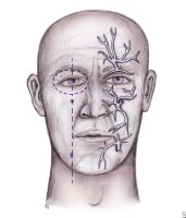
Anesthesia can be provided by topical, local, or regional block. An advantage of using regional block in the face is that the wound edges are not distorted from the local anesthetic. The areas for regional block injection are shown in the image below. Amide anesthetics (eg, lidocaine, bupivacaine, mepivacaine) are used most commonly. Allergic reactions are uncommon. When using anesthetics containing epinephrine, care should be used to avoid areas with end arteries (ie, the nose).

Distribution of nerves for regional anesthesia of Distribution of nerves for regional anesthesia of the face.

The regional block and the area of anesthesia are as follows:

Supraorbital and supratrochlear blocks – Forehead, anterior one third of the scalpInfraorbital block – Lower lid, upper lip, and lateral aspect of the noseMental nerve block – Lower lip and chinWound cleaning and irrigation

All areas should be thoroughly explored, copiously irrigated, cleaned, and débrided of devitalized tissue before closure. Irrigation lessens the risk of infection. Interestingly, regardless of irrigation, noncontaminated wounds repaired within 6 hours of injury rarely develop infection, and the overall rate of infection of repaired scalp and facial wounds is 1%.

After irrigation, gentle cleansing of the wound should be performed with a dilute povidone-iodine solution (Betadine; Purdue Pharma, LP, Stamford, Conn) or iodine solution. The wound edge (1-2 mm) can be safely removed to rid the area of devitalized tissue. Attempts should be made to make the wound edges perpendicular with the skin surface because this results in a smoother, less noticeable scar. (See the image below).

Top: Improper repair of an angled laceration. BottTop: Improper repair of an angled laceration. Bottom: Proper repair of an angled laceration, with creation of perpendicular edges for a flush repair. Repair

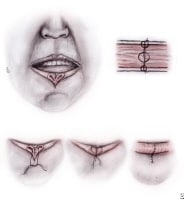
Deep wounds should be repaired in layers. Unrepaired muscle layers are much more likely to produce noticeable scarring. When performing a 2-layer closure, the deep layers should be closed with absorbable suture. Importantly, use the minimum amount of subcutaneous suture necessary because the risk of infection is related to the amount of suture used. Nonabsorbable, monofilament suture should be used for skin closure. Monofilament suture is associated with a lower risk of infection compared with a polyfilament suture. (See the image below.)

Steps to repair lip laceration. A 3-layered approaSteps to repair lip laceration. A 3-layered approach is needed, as depicted.

The suture technique should be selected based on the site of the wound and the amount of tension on the wound edges. A simple interrupted technique can be used in areas of low tension or in wounds in which the tension has been reduced with a layer of subcutaneous sutures. This technique is also useful for realigning wounds with irregular wound edges. Areas of high tension are best closed using a vertical mattress technique. All facial wounds should be repaired in less then 24 hours to decrease the risk of infection and achieve the best cosmetic result. If a delay in closure is necessary, wounds should be covered with saline-moistened gauze until the repair can be made.

Dermal adhesives, such as 2-octyl cyanoacrylate, have been shown to be equivalent to sutures for the repair of simple, clean wounds in areas of low tension.[12] The adhesives are applied topically to the wound edges. Advantages of adhesives include shorter repair time, fewer supplies, less pain during repair, and elimination of the need to remove sutures or staples at a follow-up visit. Note that dermal adhesives should not be used on the lips or mucous membranes; avoid their use in patients with poor circulation or who have a propensity to form keloids.

Staples are good alternatives to sutures in the repair of scalp lesions. Stapling involves shorter repair time and less cost compared with suture repairs. Rates of infection and inflammatory response are not higher than those associated with suture repair. During the staple application, an assistant helps to evert and approximate the wound edges, while the primary operator uses the stapler. Disadvantages include the inability to accurately align the wound edges in irregular wounds and an increased likelihood of visible scarring, thus limiting the use of stapling to the scalp.

Follow-up

The athlete should be given instructions for proper wound care, including the normal healing process and signs that might indicate the presence of complications. Anticipate any complication (eg, infection, swelling, bleeding, dehiscence) and give precise instructions for early return. The following is a list of laceration sites and recommendations on suture size and typical time to removal:

Scalp - 4-0 suture or staple, with removal in 7-14 daysForehead - 5-0/6-0 sutures, with removal in 5 daysEyebrow - 5-0/6-0 sutures, with removal in 3-5 daysFace - 6-0 suture, with removal in 5 daysEyelid - 6-0/7-0 sutures, with removal in 3 daysNose - 5-0 sutures, with removal in 3-5 daysEars - 6-0 sutures, with removal in 10-14 daysLips - 6-0 sutures, with removal in 3-5 daysPreviousNextOther InjuriesContusion

Contusions are the most common facial soft tissue injury seen by a sports medicine team. They are usually the result of blunt trauma to the face. Ice should be applied for 10-20 minutes to minimize the immediate inflammatory response. This treatment should continue for the next 48-72 hours. Over-the-counter (OTC) nonsteroidal anti-inflammatory drugs (NSAIDs) are good for symptom relief. Complications are uncommon

Abrasion

Abrasions are partial-thickness disruptions of the epidermis as a result of sudden, forcible friction. These wounds should be gently cleansed of all debris. Failure to remove all debris can lead to "tattooing" of the skin and a poor cosmetic result. Local or regional anesthetic may be required to keep the patient comfortable and achieve adequate cleaning. Lubrication of the wound using an antibiotic ointment and covering with a sterile bandage may encourage healing.

Corneal abrasion

Corneal abrasions result from loss of the surface epithelium. Disruption near the central visual axis interferes with visual acuity. Such abrasions should be treated with a course of ophthalmic topical antibiotics. Topical analgesics may be used initially, but avoid prescribing them to the athlete for home use because this may delay reepithelialization and suppress the normal blink reflex.

Emergent consultation with an ophthalmologist is warranted for suspected retained intraocular foreign bodies, and urgent consultation is needed for suspected corneal ulcerations (microbial keratitis). These injuries require close follow-up. Referral to an ophthalmologist should also be made for any athlete with continued pain after 48 hours or inadequate healing by 72 hours.

Epistaxis

Epistaxis typically does not require invasive treatment. Most often, bleeding can be controlled by maintaining continuous pressure for 10 minutes. This is achieved by asking the athlete to grasp and pinch his or her nose. While this task is performed, have the athlete tilt the head forward to avoid bleeding into the pharynx, which can lead to aspiration. Pressure should be maintained for at least 5 minutes and for up to 20 minutes. If this is unsuccessful, a second attempt should be made.

Packing the affected nostril with gauze soaked in topical decongestant may be necessary to achieve hemostasis. If the bleeding site is clearly observed, chemical cautery can be attempted using silver nitrate directly at the site. If bleeding is not controlled despite these measures, the nasal cavity should be packed from posterior to anterior with ribbon gauze impregnated with petroleum jelly. Nasal tampons may also be helpful. For particularly resistant cases, referral to an otolaryngologist may be required.

PreviousNextReturn to Play

Return to play should be based on the location and severity of the injury, sport and position requirements, and risk of the injury causing a concomitant injury. Most athletes are able to return to play immediately after treatment on the sideline or in the training room. When making return-to-play decisions, attention should be given to whether the area in question can be protected from further injury.

Previous, Sports-Related Facial Soft Tissue Injuries

Tuesday, January 28, 2014

Metatarsalgia

Background

Metatarsalgia is a common overuse injury described as pain in the forefoot that is associated with increased stress over the metatarsal head region. Metatarsalgia is often referred to as a symptom, rather than as a specific disease. Common causes of metatarsalgia include interdigital neuroma (also known as Morton neuroma), metatarsophalangeal synovitis, avascular necrosis, sesamoiditis, and inflammatory arthritis; however, these causes are often diagnosed separately. (See also the Medscape Reference articles Physical Medicine and Rehabilitation for Morton Neuroma, Surgery for Morton Neuroma, and Avascular Necrosis.)

NextEpidemiologyFrequencyUnited States

Athletes who participate in high-impact sports that involve the lower extremities commonly present with forefoot injuries, including metatarsalgia.[1, 2]

PreviousNextFunctional Anatomy

Body weight is transferred to the foot by gravity. This transfer of force is increased to the forefoot during the mid-stance and push-off phases of walking and running.[2, 3] In the forefoot region, the first and second metatarsal heads receive the greatest amount of this energy transfer. Peak vertical forces reach 275% of body weight during running, and a runner may absorb 110 tons per foot while running 1 mile.[2] Pressure studies have shown that runners spend most of the time weighted over the forefoot while running.

PreviousNextSport-Specific Biomechanics

Athletes who take part in high-impact sports that involve running or jumping are at high risk of forefoot injuries.[1, 2] Although track-and-field runners are exposed to the highest level of traumatic forces to the forefoot, many other athletes, including tennis, football, baseball, and soccer players, often present with forefoot injuries.

PreviousProceed to Clinical Presentation , Metatarsalgia

Retrocalcaneal Bursitis

Background

Pain at the posterior heel or ankle is most commonly caused by pathology at either the posterior calcaneus (at the calcaneal insertion site of the Achilles tendon) or at its associated bursae. Two bursae are located just superior to the insertion of the Achilles (calcaneal) tendon. Anterior or deep to the tendon is the retrocalcaneal (subtendinous) bursa, which is located between the Achilles tendon and the calcaneus. Posterior or superficial to the Achilles tendon is the subcutaneous calcaneal bursa, also called the Achilles bursa. This bursa is located between the skin and posterior aspect of the distal Achilles tendon. Inflammation of either or both of these bursa can cause pain at the posterior heel and ankle region.[1, 2, 3, 4, 5]

For patient education resources, see the Foot, Ankle, Knee, and Hip Center, as well as Bursitis and Tendinitis.

See related Medscape Reference topics Achilles Tendon Injuries and Tendonitis, Achilles Tendonitis, and Bursitis.

NextEpidemiologyFrequencyUnited States

Retrocalcaneal bursitis is fairly common.

PreviousNextSport-Specific Biomechanics

Inflammation of the calcaneal bursae is most commonly caused by repetitive (cumulative) trauma or overuse, and the condition is aggravated by pressure, such as when athletes wear tight-fitting shoes. Retrocalcaneal bursitis may also be associated with conditions such as gout, rheumatoid arthritis, and seronegative spondyloarthropathies. In some cases, retrocalcaneal bursitis may be caused by bursal impingement between the Achilles tendon and an excessively prominent posterosuperior aspect of the calcaneus (Haglund deformity). In Haglund disease, impingement occurs during ankle dorsiflexion.[1, 2, 3, 4, 5]

PreviousProceed to Clinical Presentation , Retrocalcaneal Bursitis

Monday, January 27, 2014

Sports Participation by Paraplegics

Overview

Competitive and recreational sporting opportunities for patients with disabilities have increased tremendously. One particular group of patients that has benefited from these opportunities and now participates in sports in ever-enlarging numbers is individuals with paraplegia. For the purposes of this article, paraplegia is defined as complete or incomplete paralysis in the lower extremities such that a wheelchair must be used as the primary mode of mobility. (See also the articles Spinal Cord Injury: Definition, Epidemiology, Pathophysiology in the Physical Medicine and Rehabilitation section of this site, and Spinal Cord Injuries in the Emergency Medicine section.)

The number of people with paraplegia continues to increase over time as general health and life expectancy has been increased to levels that are comparable to individuals without paraplegia. As a result, the demand by those with paraplegia for competitive and recreational sporting opportunities has exploded. An additional factor for the rapid growth of participation in sporting activities by those with paraplegia is the improvement in accessibility, as well as the improved designs of sporting facilities and wheelchairs, which allow for meaningful athletic competition.

Sports participation is an indispensable method of modern rehabilitation. Especially after medical rehabilitation is completed, sports have an invaluable therapeutic value in renewing the paraplegic's lost powers, helping coordination, and maintaining stamina. Today, individuals with paraplegia participate in all types of sports for competition, enjoyment, and to improve overall fitness.

NextHistory

The demand for competitive wheelchair sports traces its roots back to World War II.[1, 2] Thousands of young veterans returned from the war with physical disabilities; however, the desire to pursue sports was undiminished by these veterans' disabilities. Organized wheelchair sports began with wheelchair basketball. Teams were first formed in Veterans Administration hospitals and then expanded with community-based teams throughout the United States. Today, an extensive professional league, the National Wheelchair Basketball Association (NWBA), continues this tradition.[1] Amateur and professional leagues have formed in other team sports (eg, rugby) and in individual sports (eg, tennis, skiing, track and field).

Olympic-style games for athletes with disabilities were organized for the first time in Rome in 1960.[3] Now called the Paralympics, these games occur every 4 years for persons who fall within 1 of 6 disability groups, including spinal cord injury (SCI). A 2-week event that occurs after the regular Olympics, the Paralympics is run by the International Paralympics Committee (IPC) and has grown into an elite event that attracts nearly 4000 disabled athletes from 136 countries throughout the world.[3]

Overall, the benefits for persons with paraplegia or even tetraplegia to participate in sports activities is as varied as it is significant. Muraki and co-authors "demonstrated that sports activity can improve the psychological status, irrespective of tetraplegics and paraplegics, and that the psychological benefits are emphasized by sports activity at high frequency."[4]

PreviousNextCommonly Participated Sports

Almost any sport in which able-bodied athletes can participate in can be modified to fit participation by individuals with paraplegia. Such common sports include aerobics, air-guns, archery, basketball, bicycling/hand cycling, bowling, canoeing, fishing, football, golf, horseback riding, kayaking, soccer, quad rugby, racquetball, distance racing, rowing, sailing, road racing, skiing, sled hockey, softball, swimming, table tennis, tennis, track and field, trap/skeet shooting, water skiing, weight lifting, wheelchair fencing, and water polo.

PreviousNextSpecific Examples of More Popular SportsWheelchair basketball

The oldest team wheelchair sport is also one of the most physically demanding and one of the most popular. The NWBA nationally organizes wheelchair basketball competitions among 180 teams, which make up 21 conferences. Annual national tournaments are held for male and female divisions, junior divisions, and intercollegiate divisions.[1] The rules are modified from the National Collegiate Athletic Association rules. New wheelchair modifications continue to make the sport more competitive and even fan friendly.

Long-distance cycling/marathons

Fueled by continued technical improvements, road racing has become the most popular form of recreational and competitive activity for individuals with paraplegia. A wheelchair division now exists at almost every major marathon after the first disabled athlete competed at the Boston Marathon in 1975. The participants' racing times continue to improve and amaze. Sports 'n Spokes magazine is a publication that is dedicated to this activity and annually compiles a list of handcycle and bicycle manufacturers.

Quad rugby

Always a hit with new spectators at national wheelchair competitions, rugby is a unique sport for individuals with tetraplegia. The game is played on a basketball court by 4-member teams using a volleyball. Players are classified according to the US Quad Rugby Association (USQRA) classification system. Each class has a point value, and teams are balanced by limiting the number of points on any team to 8. The object is to carry the ball across the other team's goal line. Although the game does not really share similar rules to typical rugby, quad rugby does share a similar spirit and competitiveness. Quad rugby games can also lead to a fair amount of injuries, and the SCI physician can become quite busy during the competition.

Wheelchair tennis

First organized by the National Foundation of Wheelchair Tennis (NFWT) in 1980, the sport of wheelchair tennis is now under the aegis of the US Tennis Association (USTA). The sport follows the rules of the USTA; however, the player in a wheelchair is allowed 2 bounces instead of 1. The professional wheelchair tennis circuit is one of the few wheelchair sports that is largely independent of classification. Although this has increased the popularity of the sport among spectators without SCI, it has limited the success of players with higher-level paraplegia. On a recreational level, wheelchair tennis is one of the few sports where persons with SCI can compete with their able-bodied friends and family members.

Skiing

Also fueled by technologic revolutions, recreational and competitive skiing is extremely popular among those with SCI injuries. Persons with paraplegia and quadriplegia can ski using a sit-ski, mono-ski, or bi-ski. Numerous camps and opportunities for novices to learn and enjoy the sport are available.

Sailing

Another sporting activity that has been made increasingly accessible by continued advances in adaptive equipment is sailing. This activity offers persons with disabilities a feeling of freedom that is unmatched in almost any other sport. The National Ocean Access Project (NOAP) is a leading organization that promotes sailing for people with disabilities. The Shake-A-Leg program in Miami, Florida, operates with the support of the Miami Project to Cure Paralysis, and attracts sailors with disabilities from all over the world to test the latest equipment and train for international sailing competitions.

Fencing

Fencing can be accomplished if the wheelchair is specially modified. A fencing sports wheelchair includes a seat that is mounted to a base. A pedestal underlies the base such that the base is selectively rotatable. There is also a platform that has an upper surface to which the pedestal is affixed, as well as a lower surface. At least 3 wheels are mounted to the platform so that the wheels extend beneath the lower surface of the platform. In addition, there is a lever means for converting single-handed manual motion into forward and backward translation of the platform.

Disabled motorsports

Disabled motorsports include any motorsport that is accessible to the disabled or to wheelchair users, such as those that use motorbikes, quad bikes, go carts, etc. In fact, persons with SCI can participate in anything with an engine.

Other popular wheelchair activities:Water PoloDivingExtreme sports UK four-cross downhill mountain bikingExtreme wheelchair racing and jumping TankChair – An off-road wheelchair that is used for extreme wheelchair sportsParalympic sports

The Paralympics includes 27 sports (20 Paralympic summer sports, 5 Paralympic winter sports, 2 non-Paralympic sports) as follows[3] :

Alpine skiingArcheryAthleticsBiathlonBocciaBowls (non-Paralympic)Cross-country skiingCyclingEquestrianFootball 5-a-SideFootball 7-a-SideGoalballIce sledge hockeyJudoPower liftingRowingSailingShootingSwimmingTable tennisVolleyball (sitting)Wheelchair basketballWheelchair curlingWheelchair dance sport (non-Paralympic)Wheelchair fencingWheelchair rugbyWheelchair tennisPreviousNextParalympics

In 1948, Sir Ludwig Guttmann at the National Spinal Injuries Unit at Stoke Mandeville Hospital in Buckinghamshire, England, introduced the first Stoke Mandeville Games for World War II veterans with SCI.[2, 3] Later, other organizations were formed and sponsored competitions for persons with disabilities. As time went by, multidisability competitions developed and eventually grew into the Paralympics, or "Parallel Games for Athletes with Disabilities."[2]

After 1960, attempts were made to hold every fourth Paralympics in the Olympic host city and were successful in 1960 at Rome and 1964 in Tokyo. However, until 1988, subsequent host cities refused to host the competitions.[2] As the popularity of the Paralympics grew, the IPC was formed to coordinate the Paralympic games and other multidisability competitions on the elite sports level. In 1976, the scope of the Games was widened to accept other disabilities.[2, 3] The Paralympics games have been operating under the IPC since 1994 in Bonn, Germany. In 2000, the summer Paralympics in Sydney consisted of over 4000 athletes from 125 nations competing in 19 events (plus track and field) in both individual and team sports. For the 2004 games in Athens, over 3800 athletes from 136 nations participated.

As with any event of this size, the Paralympics faces significant challenges as it heads into the future. Many of the issues are similar to those found in the regular Olympics, including the use of performance-enhancing substances. The policy of the IPC is that the playing field should be level; unfortunately, 7 Paralympic athletes in the 2004 Athens games were sanctioned for the use of performance-enhancing agents. As a result, the IPC developed the IPC Anti-doping Code in the spirit of fair play to prevent doping in sports for athletes with a disability and in conformity with the general principles developed by the World Anti-doping Agency (WADA), the World Anti-doping Code (WADC).[3]

Another challenge is development; that is, money needs to be raised to keep the Paralympics program going. In November 2004 at the IPC Development Conference, the Paralympic Movement agreed on 5 core areas for development: athlete development, leadership development, organizational development, knowledge development, and global Paralympic development.[3]

Despite these challenges, however, there has been keen excitement regarding the 2012 Paralympic games in London, England. The list of sports and qualification guide is listed at www.paralympic.org.

PreviousNextClassification

An individual who cannot participate or compete in a sport on reasonably equal terms with persons without disabilities because of a functional disadvantage due to permanent disability is eligible for participation in that sport within the IPC program. The classification of athletes was created to equal the playing field within sporting activities in which athletes with disabilities participate. In addition to the classification systems that exist for each injury type (eg, amputation, SCI), unique classification systems exist for each sport. An athlete may be eligible for one sport and not another.

Athletes with SCI have obvious unique functional concerns. The classification of athletes with SCI, while imperfect, tries to take these concerns into account. Athletes in the Paralympics traditionally belong to 1 of 6 different disability groups, divided based on the level of the injury: amputees, persons with cerebral palsy, persons with visual impairment, persons with SCIs, persons with intellectual disability, and a group that includes individuals who do not fit into the aforementioned groups.[5] The first class, for those with cervical injury is subdivided into 3 more classes (A, B, and C) based on upper extremity strength.

The need to change functional classes is consistently reviewed. When an athlete starts competing, he or she is allocated a class that may be reviewed throughout the athlete's career. Individuals are certified in each sport to conduct the process of classification, and these officials are known as classifiers.

Certain sports rate players individually, regardless of diagnosis. For example, wheelchair basketball has a unique system; the 8 different classifications are based on sport-specific tests of shooting, passing, rebounding, pushing, and dribbling abilities, rather than a medical diagnosis or muscle function examination. Higher classification numbers represent greater basketball skills. Athletes are given a numeric point value based on their classification status; the maximum allowable points on the floor is 14.0.

In 2003, the IPC developed a classification strategy with the overall objective to support and coordinate the ongoing development of accurate, reliable, consistent, and credible sport-focused classification systems and their implementation. The IPC Classification Code Version 1.0 is a direct result of recommendations made in this strategy. The results and classification code was published in 2007 and can be found at http://www.paralympic.org/sites/default/files/document/ 120201084329386_2008_2_Classification_Code6.pdf.

The Code is complemented by International Standards that provide the technical and operational requirements for classification. The Code applies to all sports within the Paralympic Movement. Adherence to the International Standards is mandatory for compliance with the Code. The 3 International Standards are as follows:

Athlete evaluation – Procedures for the assessment of athletes and the allocation of sport classProtests and appealsClassifier training and certification

In 2011 the IPC published updated guidelines in skiing. They can be found at http://ipc-alpineskiing.org/Classification/, with 12 classification levels.

Finally, the IPC recently adopted the research paper on “IPC Position Stand – Background and Scientific Rationale for Classification in Paralympic Sport” from Tweedy and Vanlandewijck.[6]

PreviousNextMedical Benefits of Exercise

Multiple studies have found both physical and psychologic benefits from exercise in persons with SCI and that training programs for persons with disabilities can increase maximum oxygen consumption, decrease heart rate at a given work load, increase grip strength, increase arm work capacity, and increase general well-being. However, the amount of studies actually examining these issues is few, and further studies are needed. Please review some of the recent references listed in the References section.

Cardiovascular risk factors

One study showed significant association between cardiovascular risk factors and fitness, similar to that observed in persons without paraplegia.[7] Arm ergometry exercise reduced serum lipids and improved cholesterol ratios as peak oxygen consumption increased. Another study suggested that further benefit to the cholesterol profile was attained if exercise intensity achieved 70-80% of the heart rate reserve.[8]

Strength measures

Common sense would dictate that strength training increases strength and endurance. However, the extent of benefits from a coordinated anaerobic program was not definitively demonstrated until Nash et al published their findings that circuit resistance training over 4 months improved the muscle strength, endurance, and anaerobic power of middle-aged men with paraplegia while significantly reducing their shoulder pain.[9]

Cardiovascular measures

In a 1975 report, Zwiren and Ba-Or demonstrated that athletes in wheelchairs had significantly higher peak oxygen uptake than sedentary individuals in wheelchairs or sedentary individuals without SCI when performing arm exercises.[10] A 1988 study by Davis and Shephard demonstrated that highly active persons with paraplegia showed significantly better cardiovascular responses in multiple measures on cardiac function than inactive persons with paraplegia.[11]

De Groot found improvements in lipid profile, insulin resistance, and other cardiovascular parameters in patients with paraplegia who engaged in exercise.[12]

Psychosocial benefits

Persons with SCI have similar psychosocial ailments as those in persons without SCI but in larger numbers. Depression and feelings of isolation are common. Limited studies have found that exercise in persons with disabilities has led to increased feelings of well-being.[4, 13] Sports and regular activities can also encourage new friendships and help patients develop social support networks. Another study suggested that athletes in wheelchairs are more adventuresome and tough minded than inactive peers. However, whether some of these individuals participated in sports before the injury because they were more tough minded is unclear. In summary, athletes with disabilities seem to gain similar psychologic benefits from sports and exercise as athletes without SCI.

PreviousNextSpecial Medical Risks Associated With Exercise in Individuals With ParaplegiaEpidemiology

It is not entirely clear how many athletes with paraplegia become injured playing sport. One study[14] showed that 24% of all athletes participating in the 2010 Winter Paralympic Games suffered injury. The injury risk was significantly higher than during the 2002 (9.4%) and 2006 (8.4%) Winter Paralympic Games. The authors speculated this increase may have reflected improved data collection systems, but also highlighted a high risk of acute injury in alpine skiing and ice sledge hockey at Paralympic Games.

Specific injury types are discussed below.

Cardiovascular risks

Individuals with paraplegia are reported to have higher resting heart rates and lower stroke volumes while at rest and during exercise than able-bodied individuals. Additionally, persons with thoracic paraplegia have higher resting and exercise catecholamine levels than persons without SCI and persons with cervical SCI. As a result, paraplegic patients reportedly have a degree of excess cardiovascular strain, because a greater percentage of heart rate reserve is required to satisfy a given work level. Whether this translates into any risk of cardiovascular injury is uncertain.

Shoulder and musculoskeletal injuries

Persons with paraplegia depend upon the upper extremities for mobility and transfer. As a result, upper-extremity pain is the most common reported physical injury in persons with SCI, and the shoulder is commonly the most painful joint because it is often the end-bearer of weight and is subjected to significant repetitive strain. Rotator cuff tendinitis, frank tears, and general impingement syndrome are the most common shoulder injuries. Many practitioners believe shoulder pain is worsened in persons with paraplegia by muscle imbalances because overly strong anterior chest and arm muscles overpower the weak posterior scapular and cervical stabilizers. Training programs that emphasize strengthening these weak stabilizers and stretching tight anterior muscles have been proposed to reduce shoulder pain and to improve athletic performance. Other upper-extremity problems include carpal tunnel syndrome, wrist tendinitis, and elbow pain.

Autonomic hyperreflexia

Often observed when persons with SCI are exercising under the control of an electrical current, autonomic hyperreflexia (AH) is most dangerous when it goes unrecognized. Usually limited to persons with SCI above the T6 level, AH occurs because the dissociated sympathetic nervous system becomes activated and is not subject to inhibitory mechanisms. Sympathetic activity is usually initiated by an offending stimulus (eg, kinked Foley catheter). If the offending stimulus is not found, blood pressure can elevate to malignant, life-threatening levels. Some racers in wheelchairs actually induce AH as an ergonomic aid for racing, but this practice is discouraged by physicians.

Fractures

Most athletes with SCIs develop lower-extremity osteoporosis as a result of disuse, immobility, and other factors; these individuals are at higher risk for lower-extremity long-bone (eg, femur, tibia) fractures after simple falls and minor injuries. Aggressive team sports (eg, rugby, basketball) create special risks for fracture injuries. Most lower-extremity fractures in patients with paraplegia have historically been treated nonoperatively; however, in wheelchair athletes, this policy needs to be revisited because operative care might lead to a quicker return to the playing field, with better outcomes and sports achievement.

Overuse injuries

Quite common among athletes with paraplegia, such overuse can cause chronic pain syndromes. These include upper extremities tendonitis, bursitis, and skin maceration and breakdown. Cross-training, proper equipment, and proper training are all elements that lead to preventing these injuries or limiting them to some degree.

PreviousNextSpecial Topics

Wheelchair athletes require specific wheelchairs that allow for maximum function or speed. Typical measurements or design requirements that are established for the sports wheelchair include:

Removable anti-tippersAdjustable tension backrest24" wheelsAdjustable seat dumpVariable camber4" castersFore-aft axle positionRemovable bumpersHeight-adjustable footrest4 wheelsSingle anti-tipper (pivot)16" seat width and backrest heightNylon upholstery

Interestingly, Authier and co-authors published a method of sports wheelchair fabrication that costs less than USD $125 without the wheels.[15] It is these authors' hope that the publication of their design will lead to the spread of sports wheelchairs for the poor in less-developed nations.

Previous, Sports Participation by Paraplegics
Sunday, January 26, 2014

Anterior Cruciate Ligament Injury

Background

Based on statements found in the recent Orthopaedic Knowledge Update regarding the increased incidence of knee ligament injuries, the author proposes that this incidence may be associated with the current emphasis on fitness. These injuries are most often a result of low-velocity, noncontact, deceleration injuries and contact injuries with a rotational component. Contact sports also may produce injury to the anterior cruciate ligament (ACL) secondary to twisting, valgus stress, or hyperextension all directly related to contact or collision.

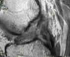
The MRI image below shows a rupture ACL:

MRI displaying a ruptured anterior cruciate ligameMRI displaying a ruptured anterior cruciate ligament.
When matched for activities, a greater prevalence for ACL injury is found in females compared with males. Approximately 50% of patients with ACL injuries also have meniscal tears. In acute ACL injuries, the lateral meniscus is more commonly torn; in chronic ACL tears, the medial meniscus is more commonly torn. The only study on the prevalence of ACL injuries in the general population has estimated the incidence as 1 case in 3,500 people, resulting in 95,000 new ACL ruptures per year.

The importance of the ACL has been emphasized in athletes who require stability in running, cutting, and kicking. The ACL-deficient knee has also been linked to an increased rate of degenerative changes and meniscal injuries. For these reasons, approximately 60,000-75,000 ACL reconstructions are performed annually in the United States.

For restoration of activity and stability, the expected long-term success rate of ACL reconstruction is between 75-95%. The current failure rate is 8%, which may be attributed to recurrent instability, graft failure, or arthrofibrosis.

Treatment options must be tailored to a patient's preoperative level of activity. The following activity levels are based on the International Knee Documentation Committee:

level I includes jumping, pivoting, and hard cutting.level II is heavy manual work or side-to-side sports.level III encompasses light manual work and noncutting sports (eg, running, cycling).level IV is sedentary activity without sports.

Nonsurgical treatment may be considered for patients who participate in level III or IV activities; all others should be considered as candidates for surgery. In addition, consider surgical consultation on any young athlete due to potential complications from recurrent instability.[1, 2, 3, 4, 5, 6]

Recent studies

A recent randomized, prospective study by Wipfler et al comparing bone-patella-bone (BTB) autografts to hamstring tendon (HT) grafts at 9 years demonstrated significantly better International Knee Documentation Committee (IKDC) scores in the HT group, with no significant differences in laxity, tunnel widening, or any other parameters.[7]

Leys et al also found equivalent IKDC scores and better long-term outcomes (radiological evidence of osteoarthritis, level of activity, knee motion and single leg hop test) in HT versus BTB in a long-term cohort study following patients for 15 years postoperatively. The HT group had higher ipsilateral graft rupture rates (17% versus 8%), but lower contralateral ACL injury (12% versus 26%).[8]

One study compared the clinical outcomes of ACL reconstruction with hamstring tendon autograft versus irradiated allograft. The results found the rate of laxity with irradiated allograft was higher than that with autograft (32.3% vs 8.3%, respectively) in the 67 patients studied. Statistically significant differences were noted between the groups in the Lachman test (P = .00011), anterior drawer test (P = .00016), pivot-shift test (P = .008), and KT-2000 arthrometer assessment (P = .00021); the anterior and rotational stabilities decreased significantly in the irradiated allograft group. No significant differences were found between the 2 groups in functional and subjective evaluations, and activity level testing; however, patients in the irradiated allograft group had a shorter operative time and a longer duration of postoperativefever.[9]

Another study evaluated the outcome of anatomic double-bundle anterior cruciate ligament reconstruction with hamstring tendon autografts in both women and men. After a 2-year postoperative evaluation, the results noted that the assessment results for ligament laxity were approximately identical in both groups.[10]

The results from another study noted that 11 years after anterior cruciate ligament reconstruction, both hamstring and patellar tendon autografts provided good long-term outcomes and stability. However, a positive result on the pivot-shift (1+) test was significantly more frequent in the patellar tendon group, as was the rate of osteoarthritis.[11]

Geib et al compared intermediate-term outcomes of ACL reconstruction by bone-patellar tendon-bone (BPTB) with the outcomes associated with quadriceps tendon with a bone plug (BQT) and quadriceps tendon without a bone plug (QT). They found that QT and BQT produced results equivalent to those of BPTB autograft in arthroscopically assisted ACL reconstruction. When compared with BPTB autograft, the quadriceps tendon autograft showed significantly better results, with less anterior knee pain (4.56% vs 26.7%), less anterior numbness (1.5% vs 53.3%), a higher percentage of arthrometer measurements showing a side-to-side difference of 0 to 3 mm (88% vs 68%), and better extension (mean loss, 0.55º vs 2.77º).[12]

According to the results of a study by Marchant et al, computed tomography is the most reliable imaging modality for evaluation of ACL bone tunnels, as proven by superior intraobserver and interobserver testing results, when compared with results obtained with MRI and radiographs. According to the authors, radiographs and MRIs were not reliable even for identifying the presence of a bone tunnel. Intraobserver kappa scores for tibial cross-sectional area using CT, radiographs, and MRI were 0.66, 0.5, and 0.37, respectively. Interobserver kappa scores for tibial cross-sectional area using CT, radiographs, and MRI were 0.65, 0.39, and 0.32, respectively.[13]

According to a study of National Football League players by Brophy et al, a history of meniscectomy, but not ACL reconstruction, shortens the expected career of a professional football player, but a combination of ACL reconstruction and meniscectomy may be more detrimental to an athlete's durability than either surgery alone. In their study, 54 athletes with a history of meniscectomy, 29 with a history of ACL reconstruction, and 11 with a history of both were identified and matched with control subjects. Isolated meniscectomy reduced the length of career in both years (5.6 vs 7.0; P = .03) and games played (62 vs 85; P = .02). Isolated ACL surgery did not significantly reduce the length of career in years or games played. Athletes with a history of both surgeries had shorter careers in games started (7.9 vs 35.1; P [14]

Lyman et al found that although ACL reconstruction appears to be a safe procedure, the risk of a subsequent operation on either knee is increased among younger patients and those treated by a lower-volume surgeon or at a lower-volume hospital. According to the authors, patients were at increased risk for readmission within 90 days after surgery if they were older than 40 years, sicker (eg, had a preexisting comorbidity), male, or operated on by a lower-volume surgeon. Predictors of subsequent knee surgery included being female, having concomitant knee surgery, and being operated on by a lower-volume surgeon. Predictors of a subsequent ACL reconstruction included age less than 40 years, concomitant meniscectomy or other knee surgery, and surgery in a lower-volume hospital.[4]

NextEpidemiologyFrequencyUnited States

An estimated 200,000 ACL-related injuries occur annually in the United States, with approximately 95,000 ACL ruptures. Approximately 100,000 ACL reconstructions are performed each year. The incidence of ACL injury is higher in people who participate in high-risk sports such as basketball, football, skiing, and soccer. When the frequency of participation is considered, a higher prevalence of injury is observed in females over males, at a rate 2.4-9.7 times greater for females.

PreviousNextFunctional Anatomy

The knee joint develops as a cleft between mesenchymal rudiments of the femur and the tibia. This occurs around the eighth week of fetal development. The cruciate ligaments appear as condensations of vascular synovial mesenchyme at the same time.

By 14weeks' gestation, the ACL and posterior cruciate ligament have divided; both have a functional blood supply, which is mainly derived from the middle geniculate artery. The inferomedial and lateral genicular arteries also provide blood supply through the fat pad.

The ACL is composed of densely organized, fibrous collagenous connective tissue that attaches the femur to the tibia. The ACL is composed of 2 groups, the anteromedial and the posterolateral bands. During flexion, the anterior band is taut, while the posterior band is loose; during extension, the posterolateral band is tight, while the anterior band is loose.

The ACL attaches to bone through a transitional zone of fibrocartilage and mineralized cartilage. On the femur, the ACL is attached to a fossa on the posteromedial edge of the lateral femoral condyle. The tibial insertion is located in a fossa that is anterior and lateral to the anterior tibial spine. The tibial attachment is noted to be somewhat wider and stronger than the femoral attachment.

The ACL is intracapsular and extrasynovial. It courses anteriorly, medially, and distally as it runs from the femur to the tibia.

The ACL receives nerve fibers from the posterior branch of the posterior tibial nerve. The main function is believed to be proprioception, providing the afferent arc for postural changes during motion and ligament deformation.

PreviousNextSport-Specific Biomechanics

The ACL is the primary (85%) restraint to limit anterior translation of the tibia. The greatest restraint is in full extension.

The ACL also serves as a secondary restraint to tibial rotation and varus/valgus angulation at full extension. Since the relationship between the tibia and femur provides little bony stability, the ligamentous structures must provide stability. When the ACL is injured, a combination of anterior translation and rotation occurs.

The average tensile strength for the ACL is 2160 N. This is slightly less than the strength of the posterior cruciate ligament and approximately half as strong as the medial collateral ligament (MCL).

PreviousProceed to Clinical Presentation , Anterior Cruciate Ligament Injury

Female Athlete Triad

Background

With the increase in female participation in sports (much of it attributable to Title IX legislation in the United States),[1, 2] the incidence of a triad of disorders particular to women—the so-called female athlete triad—has also increased.

The female athlete triad, though more common in the athletic population, can also occur in the nonathletic population. However, even though this triad was first described at the 1993 meeting of the American College of Sports Medicine (ACSM),[3, 4] associations between bone mineral density (BMD), stress fractures, eating disorders, and female athletics had been observed for decades before the syndrome was formally named.

The components of the female athlete triad, as put forth by the 1997 ACSM positional stand, consisted of disordered eating, amenorrhea, and osteoporosis.[5] Not all patients have all 3 components of the triad, and newer data suggest that even having only 1 or 2 elements of the triad greatly increases these females’ long-term morbidity.

In addition, a study by Burrows et al has suggested that the current triad components do not identify all at-risk women; rather, the authors suggest that criteria such as exercise-related menstrual alterations, disordered eating, and osteopenia may be more appropriate.[6]

Subsequent research on the female athlete triad culminated in an updated definition published by ACSM in 2007. The 2007 ACSM positional stand looks at each disorder as a point on a continuous spectrum rather than as a severe pathologic endpoint, as follows[7] :

“Disordered eating” has been replaced by a spectrum ranging from “optimal energy availability” to “low energy availability with or without an eating disorder” “Amenorrhea” has been replaced by a spectrum ranging from “eumenorrhea” to “functional hypothalamic amenorrhea” “Osteoporosis” has been replaced by a spectrum ranging from “optimal bone health” to “osteoporosis”

The 2007 ACSM positional stand also emphasizes that energy availability is the cornerstone on which the other 2 components of the triad rest.[7] Without correction of this key component, full recovery from the female athlete triad is not possible.

Often difficult to recognize, the female athlete triad can have a significant impact on morbidity and even mortality in a relatively young segment of the population. Indeed, the full impact of this syndrome may not be realized until these women reach menopause, when bone loss is accelerated.

Significant research of the triad has been ongoing, and emerging data may expand the female athlete triad to a tetrad.[8, 9] This additional research has been examining the relationship of the triad to endothelial dysfunction and the possible increased cardiac risk it athletes may face.[8, 9]

NextPathophysiologyReduced energy availability

The first component of the female athlete triad, energy availability,[7, 10] is defined as “dietary energy intake minus exercise energy expenditure” and is intended to capture those athletes who may have eating and weight concerns but do not have “significant psychopathology” and do not meet the criteria for disordered eating.

The term disordered eating itself was coined to include pathologic eating behaviors that do not meet the strict Diagnostic and Statistical Manual of Mental Disorders, Fourth Edition (DSM-IV) requirements for anorexia or bulimia; thus, it includes, but is not limited to, anorexia nervosa and bulimia nervosa.

Indeed, disordered eating includes a spectrum of behaviors ranging from simple failure to take in enough food to offset energy expenditure to preoccupation with eating and a profound fear of becoming fat (typically expressed by instituting measures such as food restrictions or the use of diet pills, laxatives, or diuretics).

Menstrual dysfunction

The second component of the triad, menstrual dysfunction,[7] describes the spectrum of menstrual function from eumenorrhea to amenorrhea and enables clinicians to capture a large portion of athletes who may have low estrogen levels but who still experience menstruation.

Menstrual dysfunction includes luteal suppression, anovulation, oligomenorrhea, and primary and secondary amenorrhea. Luteal suppression is marked by a shortened luteal phase and a prolonged follicular phase in which estradiol levels decrease. The cycle length usually does not change; the athlete will continue to ovulate—although it may be later in the cycle—and usually has regular menstruation.

Anovulation is marked by low levels of estradiol and progesterone, which deter follicular development, as well as by an absence of ovulation. Although the circulating hormone levels are decreased, female athletes will often menstruate, some experiencing shortened or prolonged cycles because of the stimulation of their uterine lining by the low levels of estradiol. Oligomenorrhea is defined as “greater than 35 days between cycles.”

Amenorrhea usually refers to secondary amenorrhea, though delayed menarche (primary amenorrhea) can occur in young athletes. By consensus, secondary amenorrhea is defined as the “absence of menstrual cycles lasting more than 3 months after menarche has occurred.” Physicians are cautioned that a full workup should be completed to rule out any other causes of menstrual dysfunction before such dysfunction is attributed to low estradiol levels stemming from low energy availability.[11]

Impaired bone health

The final component of the female athlete triad, bone health,[7, 12] describes a continuum extending from optimal bone health to osteoporosis and focuses on bone strength, which consists of BMD (or bone mineral content) and bone quality.

Bone quality refers to factors related to bone turnover rates (eg, resorption versus formation, microarchitecture or trabeculae, time for maturation of the new bone matrix, and bone geometry and size). Our current inability to measure bone quality leaves one half of the equation for bone health empty and offers an explanation for why some athletes with the same poor BMDs as their colleagues may suffer more fractures. Therefore, dual-energy x-ray absorptiometry is used as a quantitative measure of bone health.

When reporting BMD, T-scores are used for the diagnosis of osteopenia and osteoporosis. However, the T-score measures the standard deviations (SDs) below the mean to predict fracture risks for postmenopausal woman. Concern over mislabeling of our premenopausal athletes, adolescents and children, led the International Society for Clinical Densitometry (ISCD) to issue a positional stand in 2004.[12]

The ISCD’s recommendation is to determine BMD by comparing chronologic age and sex using a Z-score distribution. The Society further recommends that the term osteopenia not be used in describing bone density and that the term osteoporosis be reserved for “low BMDs” with secondary clinical risk factors such as “chronic malnutrition, eating disorders, hypogonadism, glucocorticoid exposure, and previous fractures.”[12]

Athletes with a Z-score 2 SDs below the mean are to be termed “low bone density below the expected range for age” if they are premenopausal women and “low bone density for chronologic age” if they are children. The 2007 ACSM positional stand further defined “low BMD” as “a history of nutritional deficiencies, hypoestrogenism, stress fractures, and/or other secondary clinical risk factors for fracture together with a BMD Z-score between –1.0 and –2.0” and osteoporosis as “secondary clinical risk factors for fracture with a Z-score ≤ –2.0.”[7]

Because most athletes already have a higher BMD than nonathletes, the ACSM also recommends that physicians consider performing further workup for any athlete with a BMD Z-score below -1.0, even in the absence of fracture.[7]

The bones of the lower extremities, pelvis, and vertebrae are the ones most commonly affected by poor bone health in women with the female athlete triad; stress and frank fractures of these areas are the typical manifestations. Peak bone mass is obtained between the ages of 20 and 30 years, with peak bone mineral content reached between the ages of 9 and 20 years.

Menstruating athletes gain approximately 2-4% of bone mass per year, whereas amenorrheic athletes tend to lose 2% of BMD per year. Thus, it is easy to see why athletes who are involved in high-impact sports can still be more susceptible to fractures than their nonathletic and menstruating athletic counterparts. Often these fractures are due to the increased stress sustained by these bones in the course of physical activity. In this respect, athletes with the female athlete triad are not unlike their healthy counterparts. However, those who have the triad or portions of it are more susceptible to multiple fractures, and they are also more likely to sustain fractures in larger, less commonly affected bones (eg, femoral neck, pelvis, and vertebrae).

Endothelial dysfunction

Ongoing research is looking at athletes diagnosed with the female athlete triad and the link to their endothelial dysfunction. Exact pathophysiology and etiology have yet to be established, but repeated studies have shown many athletes with the triad have endothelial dysfunction, which is a strong predictor of coronary artery health and a possible increase in atherosclerotic disease and cardiovascular event rates.[8, 9]

PreviousNextEtiology

The theory behind the female athlete triad is that this syndrome is caused by an energy drain or caloric deficit (ie, the athlete’s energy expenditure exceeds her dietary energy intake).[7, 10] This low energy availability, whether subconscious or conscious, disrupts the hypothalamic-pituitary-ovarian axis, resulting in decreased gonadotropin-releasing hormone (GnRH) pulsatility and low luteinizing hormone (LH) and follicle-stimulating hormone (FSH) levels.[13]

These changes eventually lead to decreased estrogen production, causing menstrual dysfunction. The decreased estrogen levels, in turn, affect calcium resorption and bone accretion, causing decreased bone health.

Studies have indicated that 30 kcal/kg of lean body mass is a crucial threshold for maintaining menstrual function[7] ; they have also demonstrated that increasing exercise drastically while covering the energy expenditure with increased caloric intake did not result in disruption of LH pulsatility. Conversely, decreasing an athlete’s caloric intake to less than 30 kcal/kg within 5 days resulted in decreased LH pulsatility. All of these findings support the energy drain theory.

The hormone leptin, which is secreted by adipocytes, has also garnered increased interest. Leptin appears to influence the metabolic rate, and levels are proportional to body mass index (BMI). It may be a significant mediator of reproductive function, and many studies have demonstrated that low levels of leptin correlate positively with amenorrhea and infertility. Furthermore, leptin receptors have been found on hypothalamic neurons involved in the control of GnRH pulsatility and in bone, which may also affect osteoblastic function.

Athletes in some sports that are linked to an aesthetic component or a weight class are more likely to develop the female athlete triad. These athletes often attempt to reach unrealistic weight and body fat goals dictated by their sport, to the detriment of their health.[14, 15, 16, 17]

Emotional stressors can also often be identified as inciting factors in athletes with the triad. The death of a coach or a family member, growth spurts, an illness that prevents training, and other events that an athlete cannot control often lead to disordered eating and excessive training—areas of life that the athlete can control.

For many, moving to a university setting initiates the triad cascade. Some young women move far away from family and friends, and they may carry the added responsibilities of a sports scholarship and a demanding academic workload. Collegiate athletes have the additional pressure of performing to higher competitive standards with a new coach and trainer and alongside athletes who may have had 2-3 years more experience. Not surprisingly, the prevalence of the female athlete triad suddenly increases in college freshmen.

PreviousNextEpidemiology

As noted (see above), though all female athletes are at risk for the female athlete triad or any of its components, sports that have an aesthetic component (eg, ballet, figure skating, or gymnastics) or are tied to a weight class (eg, tae kwon do, judo, or wrestling) have a higher prevalence of affected female athletes.[7, 14, 15, 18, 19]

Obtaining exact epidemiologic data is difficult because of the lack of reporting or gathering of data from athletes. Like individuals with anorexia or bulimia, many athletes with the triad try to hide their symptoms or behavior from friends, family, trainers, or coaches. This is the main reason why diagnosis is so difficult. In fact, the vast majority of cases are diagnosed only after advanced symptoms become apparent. Milder cases may be extremely difficult to diagnose if the physician does not already have a high degree of suspicion.[7, 20, 21, 22, 23]

The prevalence of low energy availability in female athletes is difficult to assess. Multiple factors (eg, the difficulty of gathering accurate caloric intake data from athletes, inability to measure energy expenditure, uncertainty regarding which sports to include or which eating attitude survey to use, and varying definitions of eating disorders) compound the issue.

It is known, however, that an athlete is at increased risk for the spectrum of reduced-to-low energy availability, with or without an eating disorder, if she has a comorbid psychological disorder, such as anxiety, depression, or obsessive compulsive disorder (OCD). In some studies, the incidence of disordered eating in the female athletic population has been estimated to be as high as 62%, with the incidence of anorexia nervosa and bulimia (as defined in DSM-IV) estimated at 4-39%.

The prevalence of menstrual dysfunction is also difficult to assess. Over a number of studies, it has ranged from as low as 6% to as high as 79%, depending on the sport studied, the patient’s age, the definition and assessment of menstrual dysfunction, the use of oral contraceptives, the training volume, and the presence of subclinical menstrual disorders, such as luteal suppression and anovulation. Studies continue to be performed, and it is hoped that more and better data will become available.

The prevalence of impaired bone health, as indicated by reduced BMD, is likewise difficult to assess because of the prohibitively high cost of DXA scans. Osteopenia has been reported to occur in 22-50% of athletes, compared with 12% of nonathletes. Osteoporosis has also been reported in 0-13% of athletes, compared with 2.3% of nonathletes. Now that the ISCD has recommended using Z-scores instead of T-scores, more research will have to be done to obtain accurate data for athletes.

Although good epidemiologic data regarding the female athlete triad are not yet available, there is reason to hope that this will change in the near future. In the meantime, many preparticipation physical questionnaires now include questions about whether the athlete is satisfied with her current weight and about how much weight she would like to gain or lose. Simple inquiries such as these may reveal the first warning signs of the female athlete triad.

PreviousNextPrognosis

For many athletes, the long-term prognosis is good. Few athletes with the female athlete triad are admitted to the hospital for inpatient treatment, and few die from their disease. However, significant long-term morbidity may affect these women later in life.

The diagnosis of the female athlete triad was established in the early 1990s, although this set of symptoms had been noted for years before it was named.[3, 16, 24, 25] However, no long-term data on future problems are available. The first generation of athletes in whom this condition was diagnosed is still years away from menopause. Thus, it is unclear whether osteoporosis occurring at a younger age affects mortality or leads to more advanced osteoporosis later in life or to an increased risk of significant fractures (eg, hip fractures).

For mild to moderate cases of the female athlete triad, some improvement in bone health is thought to occur. The lost BMD is unlikely to be replaced in its entirety, and the bone mass that should have been accumulated during this important time in bone development may or may not be regained. However, many case reports show that bone density does not increase, suggesting that losses may be permanent. Unfortunately, no long-term, double-blind, controlled studies are available (or even performable).

As more information about the female athlete triad and its complications is gathered, everyone involved may better understand the significant morbidity that can occur years or decades after the disease is diagnosed and treated.

PreviousNextPatient Education

Educating athletes may lead to earlier detection of the female athlete triad. If women know that amenorrhea is not a positive sign of hard work but a harbinger of disease, they may seek treatment sooner. Of course, the triad has a secretive nature, and by the time an athlete shows signs of disordered eating, education may not be enough to help these women seek help. If the general athletic population is aware of the signs and symptoms of this disease, the female athlete triad might be caught in its early stages.

Physicians need to do better in educating trainers, coaches, and parents (as well as the athletes themselves). These are the people who will have daily contact with the athlete, and they may be the persons who first raise concerns about a particular individual. Taking the time to talk to the athletic staff about the warning signs may help in preventing the disease or catching it in its early stages.

For patient education resources, see the Osteoporosis and Bone Health Center, the Exercise, Nutrition, and Weight Management Center, and the Women’s Health Center, as well as Anorexia Nervosa, Bulimia, and Amenorrhea.

PreviousProceed to Clinical Presentation , Female Athlete Triad

Saturday, January 25, 2014

Jumper's Knee

Background

Blazina et al first used the term jumper's knee (patellar tendinopathy, patellar tendinosis, patellar tendinitis) in 1973 to describe an insertional tendinopathy seen in skeletally mature athletes,[1] although Sinding-Larson, Johansson, and Smillie once described this condition. Jumper's knee usually affects the attachment of the patellar tendon to the inferior patellar pole. The definition was subsequently widened to include tendinopathy of the attachment of the quadriceps tendon to the superior patellar pole or tendinopathy of the attachment of the patellar tendon to the anterior tuberosity of the tibia. The term jumper's knee implies functional stress overload due to jumping (see image below).

The proximal patellar tendon is most commonly affeThe proximal patellar tendon is most commonly affected in jumper's knee.

For excellent patient education resources, visit eMedicineHealth's First Aid and Injuries Center. Also, see eMedicineHealth's patient education articles Knee Pain Overview, Knee Injury, and Tendinitis.

NextEpidemiologyFrequencyUnited States

Jumper's knee is certainly one of the more common tendinopathies affecting skeletally mature athletes, occurring in as many as 20% of jumping athletes. With regard to bilateral tendinopathy, males and females are equally affected. With regard to unilateral tendinopathy, the male-to-female ratio is 2:1.

PreviousNextFunctional Anatomy

The rectus femoris and 3 vasti muscles (ie, the vastus medialis, vastus lateralis, and vastus intermedius muscles) join in a common quadriceps tendon that inserts on the patella, the largest sesamoid bone in the human body. This same tendon is known as the patellar tendon from the inferior pole of the patella to its distal insertion at the tibial tuberosity.

Radiologic and histologic studies have shown that the posterior proximal fibers of the patellar tendon appear to be most commonly affected in jumper's knee.[2] Counter to these findings, however, biomechanical research has demonstrated that these posterior fibers can withstand greater tensile strains before failing, compared with the anterior fibers.[3]

PreviousNextSport-Specific BiomechanicsRisk factors and biomechanics

Jumper's knee is believed to be caused by repetitive stress placed on the patellar or quadriceps tendon during jumping. It is an injury specific to athletes, particularly those participating in jumping sports such as basketball,[4, 5, 6, 7, 8] volleyball,[7, 8, 9, 10] or high or long jumping.[7, 10] Jumper's knee is occasionally found in soccer players, and in rare cases, it may be seen in athletes in nonjumping sports, such as weight lifting and cycling.

Investigators have implicated sex, greater body weight, genu varum and genu valgum, an increased Q angle, patella alta and patella baja, and limb-length inequality as intrinsic risk factors.[11] However, the only biomechanical impairment prospectively linked to jumper's knee is poor quadriceps and hamstring flexibility.

Vertical jump ability, as well as jumping and landing technique, are believed to influence tendon loading.[4, 10, 12] Volleyball players with a natural ability for jumping high are at increased risk for developing jumper's knee.[13]

In a cohort of elite young volleyball players, male sex, volume of training, and match exposure were all noted to be risk factors. One third of boys aged 16-18 years developed the condition compared with 8% of girls.[14] In a cross-sectional survey of 891 nonelite athletes in the Netherlands, the prevalence of jumper's knee varied from 14.4% and 2.5% for different sports (eg, basketball, volleyball, handball, korfball, soccer, field hockey, and track and field). Younger age, taller body stature, higher body weight, and sport-specific loading characteristics of the knee extensor apparatus were all risk factors for developing jumper's knee.[15]

Overtraining and playing on hard surfaces have been implicated as extrinsic risk factors.

Interestingly, the patellar tendon experiences greater mechanical load during landing than during jumping because of the eccentric muscle contraction of the quadriceps. Therefore, eccentric muscle action during landing, rather than concentric muscle contraction during jumping, may exert the tensile loads that lead to injury.[16]

PreviousProceed to Clinical Presentation , Jumper's Knee